Adenosine Intelligence Fuels All Molecular Tags

Cells make their largest decisions at a few regulatory hubs: AMPK, which senses energy; mTOR, which decides whether to grow; p53, which guards the genome; and NF-κB, which coordinates inflammation and immunity. These hubs regulate one another in a web rather than a chain of command, but they have something in common. AMPK reads the ratio of AMP and ADP to ATP directly. mTOR checks the supply of S-adenosylmethionine through a dedicated sensor. p53 carries acetyl, methyl, ubiquitin, phosphate, and ADP-ribose tags, each delivered by an adenosine carrier, on the same few lysine and serine amino acids. NF-κB is activated by ATP-funded phosphorylation and ubiquitination inside the cell and is restrained from outside by adenosine itself. We showed how adenosine underlies the ubiquitin code.
This post shows how the other tags relate to ubiquitin and to one another through the same adenosine intelligence, the one the cell's master regulators consult before they decide.
The previous post showed that adenosine gives the ubiquitin system four things: a grip, the ink, a direction and a sensor. The grip is the adenosine monophosphate handle that makes ubiquitin’s tail reactive. The ink is the fact that nearly every kind of tag arrives on an adenosine-based carrier. The direction comes from the energy that keeps tags in place against constant removal, and the sensor is a ligase that reads ATP directly.
Since ubiquitin, acetyl, methyl, SUMO, NEDD8, phosphate and ADP-ribose are all delivered by adenosine carriers, how do these tags relate to one another? In fact, they are not separate languages. They compete, prime, read, and regulate each other. They prepare, sensitize, or precondition each other to respond differently (or more effectively) to another. Underneath all of it they draw on one shared adenosine ledger.

Competing for the Same Parking Space
Start with the simplest relation. A lysine on a protein is like a parking space, and only one car fits. The cars are acetyl groups, methyl groups, ubiquitin, SUMO and NEDD8, and which one parks changes what happens to the protein. On p53, the lysine amino acids at positions 370, 372, 373, 381, 382 and 386 can each be acetylated, methylated, ubiquitinated or SUMOylated, depending on the moment. If the enzyme p300/CBP acetylates them, p53 is protected from its inhibitor MDM2. If MDM2 ubiquitinates them, p53 heads for the proteasome to be destroyed. On histone H3, lysine 27 can be acetylated, which produces open chromatin, or trimethylated, which produces gene silencing. On IκBα, SUMO sits on lysine 21 and blocks the ubiquitin that would otherwise destroy it. At PCNA lysine 164 ubiquitin and SUMO give opposite instructions about how to deal with a damaged DNA template.
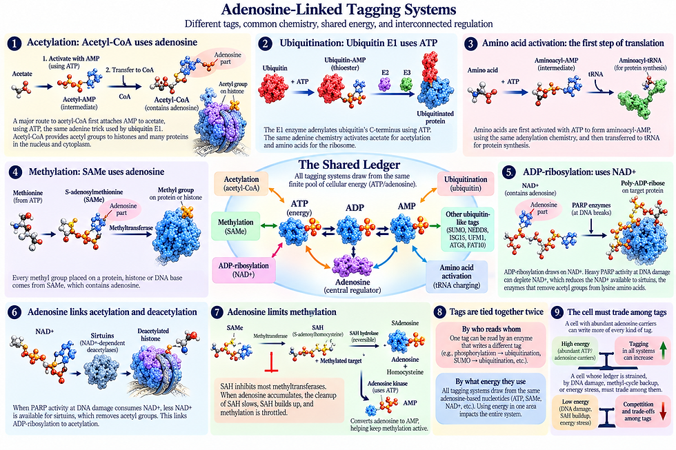
Every competitor is delivered by an adenosine-based truck. Acetyl groups come from acetyl-CoA, methyl groups from S-adenosylmethionine, ubiquitin and SUMO from ATP-driven E1 enzymes, and the sirtuins that clear acetyl groups burn NAD+. Each fight for a parking space is also a competition between the cell’s supplies of different adenosine carriers.

All, But One, Of the Tags are Fueled by Adenosine
The tags span more than three orders of magnitude in size, from a 14-dalton methyl group to an 18-kilodalton FAT10, or much larger for a long branched chain of ADP-ribose or polyubiquitin. A trimethyl group (3 methyl group) weighs almost exactly what an acetyl group does (42.05 versus 42.01 Da), yet they can have opposite effects at the same lysine amino acid on histone H3. The meaning of a tag comes from its readers, not its size.

By breadth of use, phosphorylation and ubiquitination are the most pervasive, each touching most of proteins and each with hundreds of dedicated enzymes.
By the fraction of proteins carrying it, N-terminal acetylation probably leads, at about 80% of human proteins, though it is mostly fixed rather than regulatory.
By control over protein lifespan, ubiquitin dominates, with NEDD8 as its main on-switch for the largest enzyme tag attachment, ligase, family.
By control over gene expression, methylation and acetylation of histones are central.
By clinical importance, phosphorylation (hundreds of kinase inhibitors), ADP-ribosylation (PARP inhibitors), and ubiquitin (proteasome inhibitors and targeted protein degraders) stand out. The specialized ubiquitin-like tags (ISG15, FAT10, UFM1, ATG8) are narrow in scope but essential to antiviral defense, inflammation, ER quality control, and autophagy.
There is one exception where uridine is vital rather than adenosine. But, it also interacts with adenosine in signficant ways. It is a called O-GlcNAcylation, and is a 203-dalton sugar tag that competes with phosphorylation at thousands of serines and threonine. It is delivered by a uridine carrier, not an adenosine carrier. It is the main exception among the major regulatory tags. It's complex relation to adenosine will be discussed in the next post.

Sticky Notes and Shipping Labels
You can think of the small tags as sticky notes and the large ones as shipping labels. Sticky notes are quick, cheap and easy to attach, and they record conditions. Shipping labels are heavy and commit the package to a destination.
Many pathways use a small and a large tag in sequence. A kinase, spending ATP, writes a phosphate pattern that an E3 ligase recognizes as a degron, a specific part of a protein—such as a short amino acid sequence or structural motif—that signals the cell to break down and destroy that protein. That is how IκBα and cyclin E are marked for destruction. Then the E1 enzyme spends more ATP to load the ubiquitin that the ligase attaches.
On p53, phosphorylation of the N-terminal domain at serines 15 and 20 and threonine 18 weakens its binding to its inhibitor MDM2 and helps recruit the enzyme p300/CBP, which then acetylates the C-terminal lysine amino acids. In each case a small tag sets the stage and a larger tag, or a second small tag, finishes the job. This is a recurrent pattern that is well established.

Reading One Tag to Write Another
The most striking relation is that a tag can be read by an enzyme that then writes a different tag. A ligase recognizes chains of SUMO and attaches ubiquitin to them. This is how arsenic trioxide treats acute promyelocytic leukemia: arsenic causes the PML protein to be heavily SUMOylated, enzyme reads those SUMO chains, ubiquitinates PML and sends it for destruction.
Another ligase recognizes poly-ADP-ribose, which is a polymer of adenosine-derived units built from NAD+ by PARP enzymes at DNA breaks, and ubiquitinates the proteins attached to it.
Another enzyme reads methylation on histone H3 lysine 9 and on newly copied DNA, ubiquitinates histone H3, and thereby recruits the DNA methyltransferase to copy the methylation pattern to the daughter strand.
Chromatin turns these relations into loops. The Polycomb complex writes trimethylation on H3 lysine 27, an enzyme reads that mark and writes ubiquitin on H2A lysine 119, and the ubiquitin helps recruit the enzyme back again, so each mark reinforces the other. Ubiquitination of histone H2B lysine 120 is required for methylation of H3 lysine 4 and lysine 79, which links a ubiquitin mark to methylation marks on a different histone. At DNA breaks, a repair factor binds only where it can read both a dimethylated H4 lysine 20 and a ubiquitinated H2A lysine 15, so it requires two different tags before it acts.

The Writers Wear Tags Too
The enzymes that write tags are themselves tagged. The cullin ligases, the largest family of ubiquitin ligases, are switched on when a ubiquitin-like protein called NEDD8 is attached and switched off when a protein complex removes it.
Parkin, the ligase lost in early-onset Parkinson disease, is activated when the kinase PINK1 phosphorylates a ubiquitin-like domain in Parkin, and PINK1 also phosphorylates ubiquitin itself on serine 65.
MDM2 is phosphorylated by ATM after DNA damage.
Acetylation of ubiquitin and hybrid ubiquitin–SUMO chains have also been reported.
The tagging machinery is both the author of the code and one of its subjects, which is part of what makes the system self-regulating.

A Family Sharing One Machine
Ubiquitin’s relatives are built on the same plan. SUMO, NEDD8, ISG15, UFM1, ATG8 and FAT10 all share its basic fold and are attached by the same three-step relay of E1, E2 and E3 enzymes, each with their own individualized set of three enzymes.
Humans have eight E1 enzymes, and every one of them uses ATP to adenylate the C-terminus of its tag, the same grip described in Part I. The families differ mostly in scale.
Ubiquitin has about forty E2 enzymes and more than six hundred E3 ligases, while SUMO has essentially one E2, and a handful of E3s.
SUMO is something like a small committee and ubiquitin like a parliament.
Because they share chemistry, they can occasionally cross over. Under proteotoxic stress, or when NEDD8 is abundant, NEDD8 has been reported to be conjugated by the ubiquitin machinery.

The Shared Ledger
The deepest link among the tags lies in how they use energy. While various cellular activities or marker tags appear separate, their unifying constraint is that they draw from the same finite pool of cellular energy (ATP/adenosine). All their use of energy must be balanced so that using energy in one area impacts the entire system.
Methylation is the clearest case. Every methyl group an enzyme places on a protein, histone, or DNA base comes from the universal adenosine-based methyl donor SAMe (S-adenosylmethionine). What is left over after the transfer is the breakdown product S-adenosylhomocysteine, which inhibits most transfer enzymes. This is then split into homocysteine and adenosine. This reaction is reversible and goes in the wrong direction unless both products are removed. So when adenosine accumulates, the cleanup stalls, S-adenosylhomocysteine builds up and all methylation is throttled.
Adenosine kinase, which converts adenosine to AMP using ATP, helps keep the energy situation balanced. The methyl tag thus produces adenosine as a by-product, and adenosine in turn limits the methyl tag.

Acetylation is another example. Acetyl-CoA contains adenosine, and a major route for making it from acetate, an enzyme first attaches AMP to the acetate, the same adenine trick that the ubiquitin E1 uses on ubiquitin. This has been shown to supply acetyl groups to histones in the nucleus under some conditions. The same chemical steps activates acetate for acetylation, ubiquitin for ubiquitination, and amino acids for the ribosome. ADP-ribosylation draws on NAD+, and heavy PARP activity at DNA damage can deplete it, which reduces the NAD+ available to sirtuins, the enzymes that remove acetyl groups from the lysine amino acids on which ubiquitin competes.
The tags are tied together twice. They are tied by who reads whom, and they are tied by what energy they use. A cell with abundant adenosine carriers can write more of every kind of tag, and a cell whose ledger is strained, by DNA damage, by methyl-cycle backup or by energy stress, must trade among them.
What This Does to the Idea of a Code
If the tags only competed, the system would be a list of switches. Because they prime, read, regulate and share signals with one another, it has more structure than a list. Small tags act as conditions, large protein tags act as commitments, readers translate combinations into actions, some readers deposit new tags in response, and all of them draw on the same supply of adenosine carriers.
The meaning of ubiquitin on a given protein therefore depends on its neighbors, on the competition at that residue, and on the state of the ledger. That is closer to a grammar than to a dictionary. For the argument that molecular systems are genuine agents, this is the key observation. Each enzyme has a narrow role, but the system as a whole shows conjunction, hierarchy, and feedback, which are exactly the properties we associate with decisions.
The tags together form a single combinatorial grammar.
Small tags are conditions and large tags are commitments as a general rule.
One adenosine supply couples them.
This adds up to adenosine contributing intelligence to the ubiquitin system.
The previous post showed ubiquitin’s intelligence rests on adenosine at its foundations. Here we show that the other tags do as well. This is why the tags can talk to each other. They draw on one family of carriers, they compete for the same lysine amino acids, they prime and read each other, and they regulate one another’s machinery. What looks like a set of unrelated chemical marks is better understood as one conversation conducted in many registers on a single adenosine-based foundation.

It Is The Major Hubs That Read Adenosine Activity
The tags described in this post do not act at random across all proteins. They work on a small number of the most important and vital regulatory hubs, the proteins and complexes where a cell's major decisions are made, including AMPK, mTOR, p53, and NF-κB. These hubs do not form a simple chain of command. They regulate one another back and forth. Yet they share one feature that ties them to adenosine activity: each one reads adenosine actions, and each reads a different version.
AMPK reads adenosine most literally, and its name says so: AMP-activated protein kinase—that is, adenosine mono phosphate activated protein kinase. Its γ subunit contains four repeated domains that form pockets binding AMP, ADP, or ATP. Because these adenosine nucleotides compete for the same sites, the enzyme measures not the amount of any one of them but the ratio between them, essentially the charge of the cell's battery.
AMPK spends ATP to write phosphate tags on its targets: shutting down growth by mTORC1; starting autophagy; stoping fat synthesis; and activating p53. AMPK is an adenosine sensor whose output is itself an adenosine-based tag.
mTOR reads a different version of adensine. Its complex mTORC1 decides whether the cell can afford to build, and several of its inputs are adenosine-based. When methionine and methyl donor SAM are plentiful, a binding protein releases its hold on an inhibitory complex, and mTORC1 can be active. When SAM falls, it restrains mTORC1. The methyl donor that supplies every methylation described here is also a direct input to the cell's master growth switch.
mTORC1 also senses adenosine related nucleotides through another pathway and induces an enzyme, which supplies one-carbon units for new purine synthesis. mTOR checks the methyl account and the purine account before committing the cell to growth.

p53 is the protein on which the competition of all the tag described earlier is most completely mapped. The six lysine amino acids are acetylated using acetyl-CoA and are deacetylated by SIRT1, which burns NAD+, an adenosine-based molecule. Every carrier named in this post leaves a mark on p53. Its decision whether to arrest the cell, repair it, or order its death is partly a reading of how the cell's adenosine carriers are being spent.
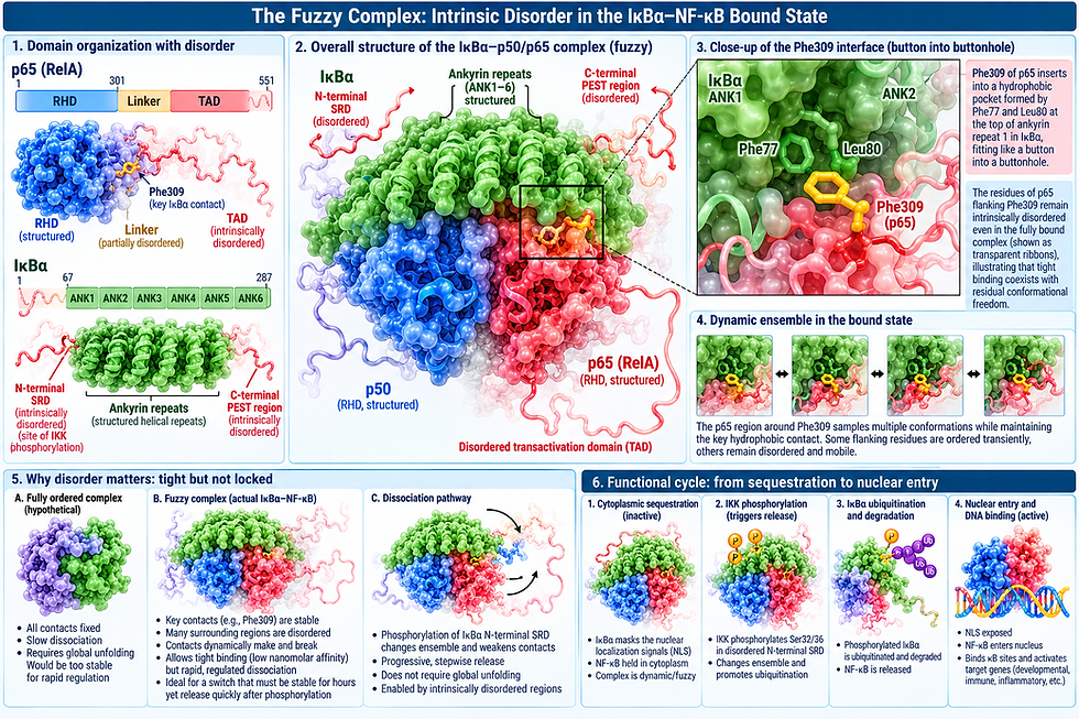
NF-κB reads adenosine in two places, inside and outside the cell. Inside, its activation follows the small-tag-then-large-tag sequence described earlier. IKK spends ATP to phosphorylate IκBα at serines 32 and 36, creating a sequence calling for destruction. In the nucleus there is competition of adenosine-based acetyl-CoA, NAD+, and SAM.
Outside the cell, NF-κB's world is governed by adenosine in its simplest forms. ATP released from damaged or dying cells is a danger signal. Acting on a receptor, it causes the second signal for the inflammasome, whose first signal, priming, is supplied by NF-κB. Then two surface enzymes, break that extracellular ATP down step by step to adenosine. Adenosine acting on another receptor raises cyclic AMP, itself an adenosine nucleotide, and suppresses inflammatory signaling. Without the receptor, it cannot shut down inflammation. The molecule that sounds the alarm, once broken down, becomes the molecule that ends it.

Adenosine At Each Layer of Architecture
The layered architecture of the vital hubs has the oldest layer, energy and nutrient sensing, which every eukaryote shares, reading adenosine as an internal ratio of nucleotides (AMPK) and as the size of the methyl and purine availability (mTOR).
The middle layer, the stress programs that guard the genome, reads adenosine as the balance among carriers competing to tag the same lysine amino acids (p53).
The youngest layer, which arose with multicellular animals to coordinate cells, reads adenosine also as a message passed between cells, ATP released as alarm and adenosine returned as calming agent.
As regulatory complexity grew, adenosine was read in new forms: first as a battery gauge, then as a set of supply accounts, then as a signal exchanged between cells.
The major hubs are best understood as readers of adenosine activity. Each layer of the regulatory architecture reads adenosine in a specific form. The layering of adenosine intelligence and activity is the way regulatory complexity emerged. The effects of adenosine intelligent activity occurs in all directions as the basis of cellular intelligent activity.