
The Dance of NF-kB
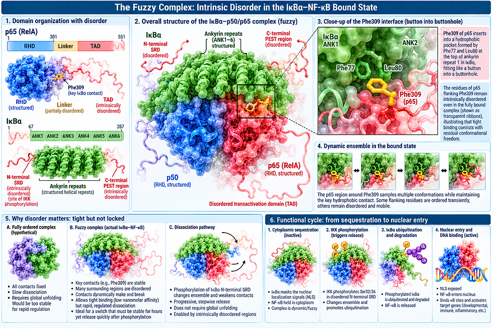
Unfolded parts of NF-κB and IκBα run the cell's inflammatory switch
The textbook story of the transcription factor NF-κB is about two folded proteins that form a pair and trigger specific region of DNA. NF-kB is most often used to refer to the very common version of NF-kB that is a pair of molecules, p50 paring with p65, from the family of 5 NF-kB molecules that form many different pairs. The name NF-kB, Nuclear Factor kappa beta, comes from the immune cell where it was first discovered.
The textbook says that an inhibitor of NF-kB, called IκBα, holds the transcription factor NF-κB in the cytoplasm. A signal arrives, a kinase marks the inhibitor for destruction, and NF-κB then enters the nucleus to switch on genes. Crystal structures support this picture, because the central pieces of both proteins do fold into well-defined shapes. But those structures leave something out that is very important. Large parts of both proteins have no fixed shape at all. These intrinsically disordered regions are not loose ends. They carry out most of the steps that decide when the system turns on, how strongly it acts, and when it turns off.

A disordered region is a stretch of protein that does not fold on its own. Instead of holding one structure, it moves continually among many shapes. Many such regions fold only at the moment they meet a partner, a process called coupled folding and binding. This flexibility has practical advantages. A disordered segment is exposed, so enzymes that add or remove chemical tags can reach it easily. It can stretch across a distance to touch a surface a folded domain could not reach. It can make many weak contacts at once and combine the effects. And it can switch between states quickly, which makes it well suited to controlling timing.
The system's start button is disordered. The first seventy or so amino acids of IκBα form a flexible tail containing two serines, serine 32 and serine 36. This is the molecule that restrains NF-kB and when it is released NF-kB is activated. When the kinase IKKβ is activated, it phosphorylates these two serines, and the phosphorylated tail is then recognized by the ubiquitin ligase βTrCP, which tags IκBα for destruction by the enzymes that degrade proteins. Because the tail is already exposed and mobile, nothing has to unfold before the kinase can act on it. This is one reason the response can begin within minutes of a signal.
The inhibitor's lifespan is controlled by disorder. IκBα's core is a stack of six ankyrin repeats, a type of chemical Velcro that holds only NF-kB to inhibit it. The last two repeats, the fifth and sixth, are only loosely folded when IκBα is free. They flicker between folded and extended forms. In that state the free protein is quickly consumed by the enzyme that degrades it, even without a ubiquitin tag that signals the molecule for destruction, and it survives only a matter of minutes in cells. When IκBα binds NF-κB, those two repeats fold firmly, and the bound inhibitor survives for hours. The act of binding decides whether the inhibitor lives or dies. As a result, IκBα accumulates only as far as there is NF-κB to hold. Excess inhibitor is cleared continuously, and once a signal destroys the bound pool, it cannot quickly be rebuilt.

The inhibitory complex itself is unusual. The two proteins bind extraordinarily tightly, yet parts of the interface stay in motion. The nuclear localization segment of NF-κB's p65 subunit forms a short helix that packs against the first ankyrin repeats of IκBα, while neighboring stretches remain flexible even in the assembled complex. Biophysicists call such an arrangement a fuzzy complex. This means that this residual looseness lets the complex be held tightly and yet remain open to rapid rearrangement when the next step of the cycle arrives.

That next step is the shutoff. When NF-κB is active in the nucleus, one of the genes it switches on is the gene for IκBα. The newly and rapidly made inhibitor enters the nucleus and removes NF-κB from its target genes far faster than NF-κB would leave DNA on its own. This is called molecular stripping where IκBα briefly forms a three-part complex with NF-κB and DNA. The folded ankyrin repeats grip one surface of NF-κB, while IκBα's disordered and strongly negatively charged C-terminal region, the PEST sequence, reaches toward the positively charged surface that holds DNA. There it competes with the DNA's negative charges and twists the NF-κB dimer so that the DNA lets go. Termination is an active process performed by a flexible tail, not a passive wait for NF-κB to drift away.
P105

Disorder also explains how the ancient precursor proteins p105 and p100 give rise to working transcription factors rather than being destroyed. Between their DNA-binding domain and their ankyrin-repeat tail lies a disordered, glycine-rich region of low sequence complexity. When the enzymes degrade these precursors, its motor grips this region poorly and slips. Next to the glycine-rich region sits the tightly folded DNA-binding domain, which the slipping motor cannot pull apart. Processing stops, and the intact front half is released as p50 or p52. Without the glycine-rich region, the whole protein is simply degraded. The oldest release mechanism in the NF-κB family depends on a stretch of protein that has no structure at all.

On the activating side, most of the p65 subunit beyond its folded core is disordered: a long flexible linker followed by the transactivation domain, the region that recruits the machinery for turning genes on. The linker lets the activation domain swing freely, reaching partner proteins regardless of how the DNA-binding core sits on DNA. Within the activation domain, two short segments, TA1 and TA2, form helices when they bind the coactivators CBP and p300, the same coupled folding used by p53 and many other transcription factors. Phosphorylation adjusts this recruitment. Serine 276, in the folded core, is phosphorylated by kinases such as PKA and MSK1 and promotes CBP/p300 binding. Serine 536, in the disordered activation domain, is phosphorylated by IKKs and other kinases and enhances activation. Because these sites are exposed, p65's output depends on which kinases have recently acted in the cell, so the molecule integrates several signals rather than responding to one.
The activation domain also affects how p65 binds DNA in the first place. It increases the p65/p50 dimer's overall affinity for DNA while reducing how sharply it distinguishes between DNA sequences, the opposite of what one might expect. One proposal is that looser, broader DNA contacts help the dimer search the genome for its sites more efficiently.

Disordered regions may also help build higher-order structures at active genes. At super-enhancers driving strongly induced inflammatory genes, p65 works with BRD4, which binds acetylated p65, together with the Mediator complex and RNA polymerase II. These factors are thought to gather into dense, droplet-like hubs, or condensates, held together by the many weak contacts that disordered regions make. Such hubs would concentrate the transcription machinery and speed up gene activation. Stripping by IκBα would also dissolve the hub by removing p65.

Taken together, these findings change how the whole system should be understood. In many cells, nuclear NF-κB rises and falls in oscillations with a period of roughly ninety minutes to two hours.The system is operating under kinetic control: at any moment the amount of active NF-κB is set not by a simple balance between bound and unbound states but by the rates of synthesis, degradation, folding, and stripping. Several of those rates run through disordered regions: the exposed start signal of IκBα, the loosely folded repeats that set its lifespan, the PEST tail that performs stripping, and the activation domain that drives production of new IκBα. Change any of them, and the timing of the response changes. Not every step depends on disorder, since the upstream activation of IKK relies on folded kinases and ubiquitin scaffolds, but the steps that set the tempo largely do.

The folded domains of NF-κB and IκBα supply the system's recognition: which DNA sequence, which partner, which surface. The disordered regions supply its timing and its conditions: how fast it switches on, how long it stays on, how it is switched off, and which signals shape its output. Disorder also offers a convenient control. Because a disordered region has no structure to break, changes in its sequence can adjust a rate without damaging the protein's fold, letting timing be tuned gradually. In this sense, much of the system's intelligence lives in its unfolded parts. The structure knows what to bind. The disorder knows when. As with RNA virus, the intelligence is in the rapid interactions of the disordered regions.


