Intelligence in Deciding Where the Cell Takes Out Trash
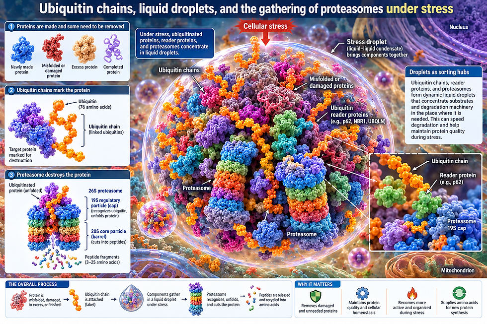
Ubiquitin chains, liquid droplets, and the gathering of proteasomes under stress
Every cell is constantly making proteins and constantly getting rid of them. Some proteins are damaged, some are misfolded, some were made in excess, and some have simply finished their jobs. To dispose of them, the cell marks each one with a small protein called ubiquitin, 76 amino acids long. One ubiquitin is often attached to another, and another to that, building a chain. The chain is a label. The machine that reads the label and destroys the protein is the proteasome, a barrel-shaped complex that unfolds its target and cuts it into short pieces.
For a long time this was pictured as a scattered process: tagged proteins drifting until they bumped into one of the many proteasomes spread through the cell. A newer picture adds something surprising. Under certain conditions, the tags, the proteins that read the tags, and the proteasomes themselves gather into small droplets, much as oil gathers into drops in vinegar. These droplets bring the disposal machinery together in the place where it is needed.

Droplets in Cells
Biologists call these droplets condensates. They have no membrane around them. They form because certain molecules have many weak, sticky contact points, so each molecule can hold onto several others at once. Biochemists call this multivalency. When enough such molecules are present, they stop mixing evenly with the surrounding fluid and separate into a denser drop, a process called phase separation. The drop stays liquid: molecules move in and out, and two drops can fuse into one. Anything that adds sticky points pushes molecules into the drop. Anything that occupies or hides those points dissolves it.
Ubiquitin chains are well suited to this. A chain of four ubiquitin molecules offers four binding surfaces, mainly a small greasy patch centered on the amino acid isoleucine 44. A protein that binds ubiquitin can therefore be linked to several others through a single chain, and a network can form.

Not All Chains are The Same
Ubiquitin has seven lysine amino acids, plus its first amino acid, where the next ubiquitin can be attached. The attachment point decides the chain's shape, and the shape carries meaning.
Chains linked through lysine 48, called K48 chains, are the classic signal for destruction by the proteasome. They tend to fold up compactly, with neighboring ubiquitin molecules pressing their sticky patches against each other, so fewer patches are exposed.
Chains linked through lysine 63, called K63 chains, are more often signals for repair, signaling, or disposal by autophagy, the cell's other route of destruction, in which material is wrapped in a membrane and digested. K63 chains are stretched out and open, with their sticky patches exposed.
The same building block, joined at a different point, produces a different object that presents a different face to the proteins that read it.

A Reader That Ubiquitin Dissolves: Ubiquilin-2
Ubiquilin-2 is a ubiquitin reader (or ubiquitin receptor) that recognizes and binds to a ubiquitin molecule that is already attached to a target protein and then determines the targets fate. Ubiquilin-2 contains a specific domain architecture that allows it to bridge two separate parts of the cellular cleanup process.
It has a reader end that recognizes and binds to a protein that is already tagged with a ubiquitin chain that needs to be destroyed. Then, its shuttle end physically binds it directly to the proteasome (the cell's garbage disposal). By reading the code on the target protein and physically walking it over to the proteasome, it successfully shuttles damaged or misfolded proteins out of the cellular environment.
In between are stretches that let ubiquilin-2 stick loosely to other copies of itself, including a proline-rich region where many mutations that cause ALS, the paralyzing neurodegenerative disease, are found.
Because of its ability to bind to copies of itself, biquilin-2 can increase local molecular density and form liquid droplets (also called phase separated condensates) inside the cell. When ubiquitin binds to ubiquilin-2, the droplets dissolve because ubiquitin occupies regions that ubiquilin-2 would otherwise use to hold onto its neighbors. When ubiquilin-2 enters stress granules, the droplets cells form when stressed, binding ubiquitin breaks up the connections and draws ubiquilin-2 it back out of the droplet (granule).
The type of chain that ubiquitin forms matters. K48 chains dissolve ubiquilin-2 droplets. K63 chains strengthen the droplets because the open K63 chain works as a bridge between several ubiquilin-2 molecules binding them together and continuing to create the density of molecules that forms the droplets. So, the same molecular reader can be pushed into a droplet or out of one depending on which kind of chain it meets. The ALS mutations change how readily ubiquilin-2 forms droplets and make it more likely to harden from liquid into solid clumps, which links this physical behavior to disease.

A Reader That Ubiquitin Gathers: p62
The protein p62 works in the opposite direction. One end of p62 links to other p62 molecules end to end, forming long helical filaments. The other end binds ubiquitin, preferring K63 chains. Ubiquitin chains added to the filaments increase the local molecular density and drive p62 into droplets, which are called p62 bodies. Because a single polyubiquitin chain has multiple ubiquitin units, different units on the same chain can bind to the domains of different p62 molecules simultaneously. By holding separate p62 proteins together at their ends, the polyubiquitin chains act as a physical molecular bridge. When you combine this bridging effect with the fact that p62 naturally links to itself to form long filaments, the proteins cross-link into a dense, sticky network.
This collective "stickiness" causes the molecules to condense and separate out of the cellular fluid into liquid-like droplets known as p62 bodies. Here two kinds of stickiness add together: the filaments of p62 provide one, and chains bridging the ubiquitin-binding ends provide the other. These bodies mostly lead tagged material toward autophagy rather than the proteasome.
The cell adjusts p62 bodies by attaching phosphate groups. Enzymes attach phosphate on serine 403, which makes p62 grip ubiquitin more tightly. Phosphate on serine 349 lets p62 capture a protein, which releases a factor to switch on the cell's defenses against oxidative damage. This signals to another protein, which helps the bodies form. In p62, then, the droplet is not only a disposal site but also a switch that sends signals.

Proteasomes Gather
With high cell concentrations, proteosomes themselves entered droplets. Forming these droplets required ubiquitin-tagged proteins. It also required a shuttle protein that like ubiquilin-2, has one domain that docks on the proteasome, but it has two ubiquitin-grabbing domains instead of one. The shuttle protein holds the proteasome with one end and ubiquitin chains with the other, and chains of about four or more ubiquitin molecules provide enough contact points to knit many such bridges into a network. The drops also contained p97, a machine that pulls tagged proteins out of complexes and unfolds them for the proteasome.
The tagged proteins inside these drops turned were largely ribosomal proteins that had failed to join ribosomes, building blocks left without a structure to join. The drops dissolved as these proteins were destroyed, because the drops are places of active disposal rather than places of storage.
Another type of drop in the nucleus forms containing p62, proteasomes, and tagged proteins. In this droplet, proteins are actively broken down in the nucleus. This shows that disposal is not limited to the autophagy route using lysosomes in the cytoplasm. Also, when cells have few nutrients, proteasome drops form in the nucleus. These drops depend on p62 and are linked to signals that lead to cell death. In this situation, ubiquitin serves signaling rather than faster disposal.

The Same Move Can Mean Different Things
Gathering proteasomes together does not always mean speeding up disposal. When the cell runs out of glucose and stops growing, their proteasomes leave the nucleus and gather into proteasome storage granules in the cytoplasm. These protect the proteasomes from being destroyed by autophagy while the cell waits, and the droplets come apart when food returns letting out the proteosomes. Gathering here means pausing and protecting, not working faster.
Droplets also appear at the step of tagging. Part of an enzyme complex that attaches ubiquitin to proteins forms droplets together with the proteins it targets, so that the tagging happens inside the droplets. Blocking droplet formation also slows the destruction of its targets. Ubiquitin also governs how stress granules end. Stress granules that form without ubiquitin tagging, and without destroying the proteins, after heat stress, they cannot come apart without it. Tagging of the granule protein, followed by p97 pulling it out, takes them apart. While ubiquitin is conventionally thought of as a "destruction tag" that marks damaged or misfolded proteins to be broken down by the proteasome, this specific mechanism acts differently.
The broader idea is that cells form droplets as a general strategy for bringing disposal machinery together on demand, and that the liquid nature of the droplets matters.

The Cell Decides
A single reader protein treats a K48 chain and a K63 chain as instructions to take up opposite physical states, scattering in one case (breaking up a droplet) and gathering in the other (forming or staying in a droplet). And when a cell is suddenly shocked, it does more than tag the proteins that went wrong. It rearranges the space where those proteins are destroyed, pulling the destroyers, the shuttles, the unfolding machine, and the targets into one place, then letting them go when the it is safe to do so.
The cell decides where to work. The mechanism involves weak sticky contacts, chain shapes, and bridging shuttle reader molecules. The decision emerges from how many small molecular choices to either bind or let go add up, and from the fact that the shape of a ubiquitin chain carries information that changes the physical organization of the cell. This is a form of molecular intelligence in the distributed sense with readers, labels, and rules of attraction, which together put the machinery where it is needed. This intelligence determines the outcome and triggers the needed activity.