
The Life of mTOR : the Cell's Coordinator of Growth, Energy, Construction, and Survival
Updated: 6 days ago

mTOR Decision-Making System
A Molecular System That Makes Decisions
Among the thousands of molecular systems operating inside a human cell, few coordinate as many different activities as mTOR. Its name, mechanistic target of rapamycin, makes it sound like simply another enzyme. Technically, mTOR is a protein kinase: it transfers phosphate groups to other proteins and thereby changes their behavior. But describing mTOR merely as a kinase is like describing a brain as a collection of electrically active cells. It identifies the physical mechanism while missing the extraordinary level of organization produced by it.
mTOR sits near the center of an immense network of cellular communication. It continually receives information about nutrients, energy, oxygen, growth factors, hormones, cellular damage, stress, and the condition of cellular organelles. It integrates these signals and changes the behavior of numerous downstream systems accordingly. Through these actions, it helps determine whether a cell should grow or conserve resources, manufacture proteins or recycle them, build membranes or break down stored material, proliferate or remain quiet, and invest in immediate growth or long-term maintenance.
Its decision-making emerges from molecular interactions, phosphorylation reactions, feedback loops, and localization. Its behavior has many characteristics we ordinarily associate with intelligence: gathering information, comparing competing conditions, setting priorities, coordinating many activities toward a common objective, detecting changing circumstances, and reversing strategy when conditions change.
Two Interconnected Command Systems
Much of mTOR's versatility comes from the fact that the large mTOR protein operates as part of two different molecular assemblies, mTOR complex 1 (mTORC1) and mTOR complex 2 (mTORC2). The same central kinase participates in two regulatory systems with substantially different interacting responsibilities.
mTORC1 is primarily concerned with the relationship between resources and growth. It asks whether the amounts of amino acids, energy, oxygen, and growth signals are sufficient to justify cellular construction. When conditions are favorable, it promotes construction by stimulating protein synthesis, ribosome production, lipid synthesis, nucleotide production, and reprograms how the body processes glucose to use it as physical building blocks for cellular construction. It stimulates cellular growth while suppressing all recycling programs including autophagy — the highly regulated survival mechanism where a cell intentionally breaks down its own damaged proteins, worn-out organelles like old mitochondria, and cellular debris, recycling them into raw molecular building blocks or energy.
mTORC2 is important for cellular survival, metabolism, membrane organization, and the cytoskeleton. Through a series of vital kinase pathways it influences whether cells survive, how they respond to insulin, how their membranes behave, and how they establish shape, polarity, and movement. A major pathway mTORC2 triggers is one of the cell's main growth and survival switches — a kinase called AKT. The name AKT comes from the way it was discovered; its other name, related to what it actually does, is Protein Kinase B.

When nutrients and growth factors are available, AKT tells the cell to grow, make proteins, use glucose efficiently, and avoid programmed cell death. A second kinase, SGK, is a backup and partner to AKT that helps promote cell survival, growth, and ion transport. It can perform many of the same jobs, especially when AKT activity is reduced, making it important in normal physiology and many cancers. A third, PKC, is a family of signaling enzymes activated by lipid signals and by calcium. PKC helps cells respond to external signals by regulating secretion, gene activity, cell shape, movement, and communication with neighboring cells.
The two mTOR complexes are assembled using different partner proteins that determine which targets each complex acts on and how each complex is regulated. For each complex, mTOR associates with a primary scaffold protein that organizes the vast number of molecules it contacts. mTORC1 is built around a protein called Raptor, which functions as a target recruiter and determines where in the cell the complex is active. mTORC1 also includes another subunit, mLST8, which stabilizes the reactive domain of mTOR, and two negative regulators — PRAS40 and DEPTOR — that sit within the complex and restrain its activity until positive signals override them.
mTORC2 is built instead around a protein called Rictor, also accompanied by mLST8, and by a protein called SIN1 that comes in four alternatively spliced forms and connects the complex to the Ras signaling network. Ras comes from the animal in which it was discovered. It is a vital cellular mechanism that transmits extracellular signals from surface receptors to the nucleus, controlling cell growth, division, survival, and differentiation. The practical consequence of this architecture is that the two complexes, though sharing the same catalytic kinase, respond to different signals, phosphorylate different targets, and perform different cellular functions — meaning the cell has, in effect, two intelligently coordinated control centers built around the same enzymatic core.
The distinction between the two complexes was initially defined by using the molecule rapamycin, which binds to a small protein forming a rapamycin complex that docks on a specific region of mTOR called the FRB domain, blocking access to mTORC1's targets. Rapamycin cannot dock on mTOR when it is already assembled into the mTORC2 configuration. Most of what is known in molecular detail about mTOR biology has been learned through using rapamycin as a tool, which is why our understanding of mTORC1 is considerably deeper than our understanding of mTORC2.
These two mega-complexes are not isolated systems. They are embedded in feedback circuits connecting nutrients, receptors, metabolic pathways, organelles, and transcriptional regulators. The result is not simply an on/off switch but a continuously adjustable control network.

Gathering Information About the Cell and Its Environment
Before committing resources to growth, mTORC1 receives information from a remarkable variety of sources, one being specialized sensors and signaling systems that ultimately influence enzymes on the lysosome surface that attract mTORC1 to the membrane.
A major location for mTOR sensing is organized around the lysosome, the cell's digestive organelle, a membrane-enclosed compartment packed with enzymes that break down proteins, lipids, and nucleic acids. It is one of the primary locations at which mTORC1 measures the cell's amino acids, lipids, and sugars. These three mostly dictate whether mTORC1 physically anchors to the lysosomal membrane, while nucleotides dictate whether the complex actually turns on.
On the surface of the lysosomal membrane sits a multiprotein complex that ties a pair of enzymes to the lysosomal surface. When nutrients are present in the cytoplasm, signals are fed through a regulatory cascade. When satisfied, the enzymes tied to the lysosome shift into an active configuration that physically summons mTORC1 to the lysosomal surface.
Once docked at the lysosome, mTORC1 can be activated by another, separate input: a signal from a growth factor. There are multiple growth factors involved, but one is the well-known insulin. Growth factors activate a signaling cascade beginning with a kinase, which generates a lipid messenger at the cell membrane. This activates the other famous kinase, Akt, which sends a series of messages that ultimately activate an enzyme already tied to the lysosome surface. mTORC1 is fully activated only when nutrient sufficiency and growth factor signals both indicate a favorable environment — either signal alone is not sufficient. The cell requires confirmation on two independent dimensions before committing to full molecule-building activity.
At the same time, mTOR receives another stream of information from outside the cell. Insulin and other growth factors activate receptor pathways involving AKT and a lipid signal pathway. These signals communicate something very different from the nutrient sensors: they indicate that the organism is providing a physiological environment favorable for growth.
Energy status provides another input. A cell might have abundant amino acids but insufficient ATP to undertake expensive biosynthesis. The energy sensor AMPK detects such circumstances and restrains mTORC1, both directly and through other upstream regulators. The cell therefore does not respond simply to the presence of building materials; it also asks whether it can afford to use them.
Oxygen availability, glucose, oxidative conditions, DNA damage, ER stress, and other forms of cellular stress feed into the network as well. The striking feature is integration. No single signal necessarily determines the outcome. Numerous pieces of information converge on the same regulatory machinery.
The system behaves as though it were asking: What resources do I have? How much energy is available? What signals am I receiving from the organism? Is the environment safe for growth? Is the cell damaged? What should be done now?
What emerges from this catalog of inputs is not a simple on/off switch but a genuine multi-dimensional integrator. mTOR does not respond to amino acids alone, or energy alone, or growth factors alone. It integrates all of them simultaneously, weighting their contributions to arrive at a composite assessment of whether the cell is in a condition to grow. No single favorable signal is sufficient to fully activate it; no single unfavorable signal is sufficient to fully suppress it if others are positive. This is a computational function — and it is performed not by neurons or circuits but by interacting proteins at the lysosomal surface.

II. The Decision to Build
When the combined information favors growth, mTORC1 initiates a coordinated building program. A growing cell needs proteins, which requires ribosomes with ribosomal RNA and ribosomal proteins. New cellular structures require membranes, which require lipids and cholesterol. Cell division requires DNA and RNA, which require nucleotides. All of this requires enormous amounts of ATP and metabolic raw material. mTOR coordinates all of these demands.
Protein Synthesis and Ribosome Production
To stimulate protein synthesis, mTORC1 phosphorylates a series of proteins, which changes the machinery controlling protein production. With another series of phosphate tags, cap-dependent protein production is initiated. As a result, the cell's protein-production machinery becomes more active.
mTOR promotes production of more ribosomes. It influences production of ribosomal RNA, ribosomal proteins, and components needed to assemble functional ribosomes. Through effects involving cellular RNA polymerases I, II, and III and their regulatory machinery, mTOR increases the cell's capacity to manufacture proteins on a large scale.
Ribosome synthesis — the manufacture of the ribosomal machinery itself — is also under mTOR's direct control. mTORC1 tags an enzyme and starts a cascade of signals that drives RNA polymerase I to produce the ribosomal RNA that forms the structural core of ribosomes. This additionally promotes transcription of ribosomal proteins and other factors related to protein production. In a separate pathway, mTORC1 stops a repressor of RNA polymerase III, unleashing Pol III to produce the transfer RNAs used during protein production.

Lipid Metabolism
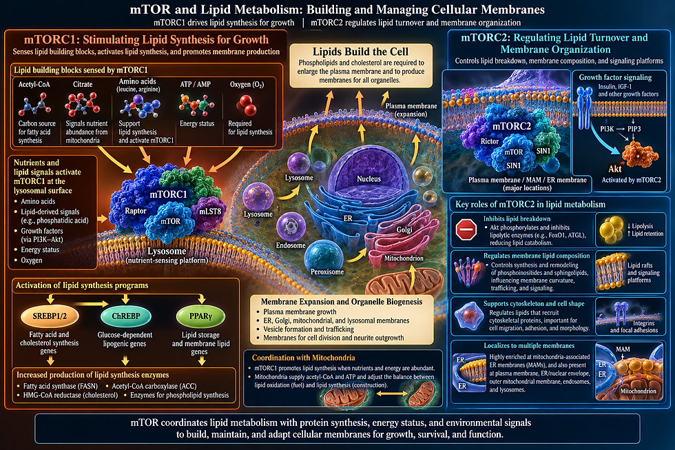
Protein production is only part of cell growth and construction. mTORC1 stimulates lipid metabolism through pathways controlled by master transcription factors. Fatty acids and cholesterol are required to produce and expand cellular membranes. A growing cell must enlarge its plasma membrane and produce membranes for the ER, Golgi, mitochondria, lysosomes, and other structures.
Lipids present a particular challenge because the cell cannot grow without membrane. Every new organelle, every vesicle, every dividing cell, every expanding dendrite requires phospholipids and cholesterol in the right proportions at the right time. mTOR reads the availability of lipid building blocks directly, sensing the levels of acetyl-CoA and citrate, the raw materials from which fatty acids are made, as well as the energy molecules ATP and AMP and the availability of oxygen.
When conditions are favorable, mTOR activates the master transcription factors for fatty acid and cholesterol synthesis, increases production of the enzymes that carry out these reactions, and coordinates with mitochondria to balance how much lipid is being burned for fuel against how much is being saved for construction. During active growth it specifically promotes synthesis of the phospholipids that form the membranes of new organelles.
The mTORC2 cluster contributes a complementary role: through its activation of Akt, it inhibits the breakdown of lipids and regulates the curvature and composition of membranes, including the specialized lipid rafts that serve as docking platforms for signaling proteins and cytoskeletal anchors that define cell shape and movement. Sphingolipid and phosphatidylinositol metabolism — two lipid classes central to how cells signal to each other and to their own interior — are tuned by mTORC2 as part of this same membrane management program.
Nucleic Acid Synthesis
Nucleic acids are another requirement that cannot be ignored. Rapidly growing cells need more ribosomal RNA, more transfer RNA, and more messenger RNA to keep the protein factories running, as well as nucleotide building blocks for DNA when the cell prepares to divide. All of this requires a ready supply of amino acids, ATP, and carbon, and mTOR monitors these prerequisites before committing to nucleic acid synthesis.
When conditions are right, mTOR activates the enzymes that produce both purines and pyrimidines, and it simultaneously activates RNA Polymerase I in the nucleolus to transcribe ribosomal RNA and RNA Polymerase III to produce transfer RNAs and other ribosomal RNA components. It increases expression of the proteins and assembly factors that turn these raw RNA molecules into functional ribosomes. The result is a coordinated upswing in the cell's entire nucleic acid network, timed precisely to when there is enough energy and material to sustain it. When AMPK signals that fuel is running low, all of this is shut down promptly — nucleotide synthesis stops, RNA transcription contracts, and the cell conserves what it has.

Glucose Metabolism
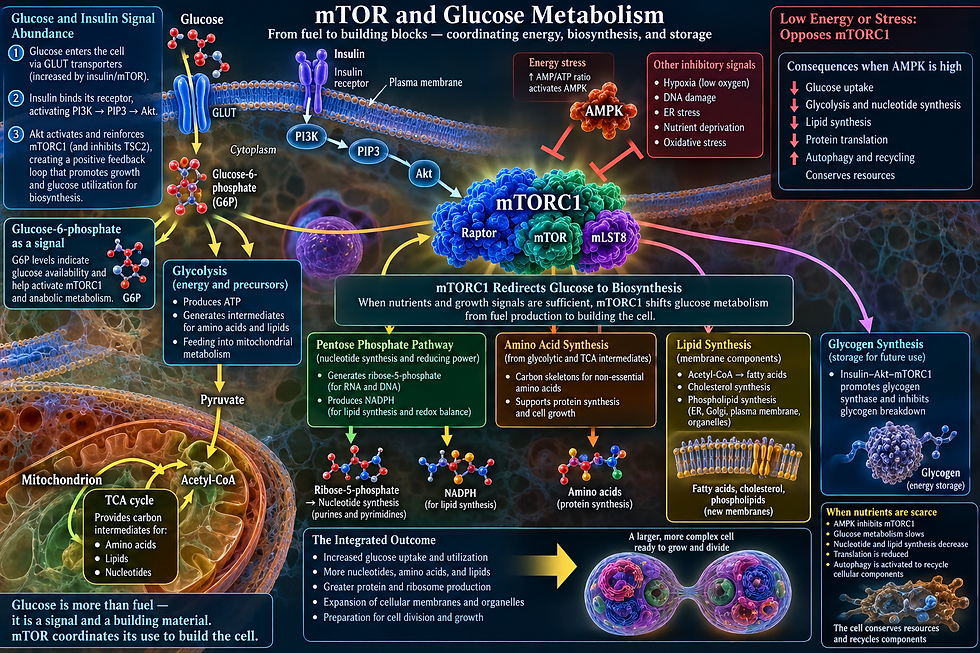
Glucose metabolism is reorganized as well. Carbon obtained from nutrients can be directed through glycolysis and related metabolic pathways not simply to produce ATP, but also to generate precursors for amino acids, lipids, and nucleotides. The cell's metabolism becomes oriented toward construction.
mTOR treats glucose not just as a fuel but as a signal and a building material. When glucose and insulin are present, lipid signaling cascades activate Akt, which in turn reinforces mTORC1, creating a positive feedback loop that accelerates the entire building program. Sensing glucose-6-phosphate as an indicator of glucose availability, mTOR triggers increased glucose uptake and redirects the flow of sugar through metabolism, not just toward energy production through glycolysis but toward the pentose phosphate pathway, which generates the ribose-5-phosphate needed for nucleotides and the NADPH needed for lipid synthesis. Glycogen synthesis is also promoted through the insulin-Akt axis, storing glucose for future use.
The essential shift mTOR engineers when nutrients are abundant is from burning sugar as fuel to using sugar as raw material for building the cell. When fuel falls and AMPK rises, mTOR reverses course entirely — glucose production slows, translation stops, and the cell turns toward recycling and autophagy to extract what it needs from its own components.
An Integrated Metabolic Program
mTOR produces an integrated metabolic program in which lipids, nucleic acids, sugars, and proteins are all monitored and produced together, in proportion, with each branch of synthesis feeding information back into the others. Membranes are built as organelles are assembled. Ribosomal RNA is made alongside the ribosomal proteins that will use it. Nucleotides are synthesized in step with the demand from active ribosomes. Glucose is redirected toward construction rather than combustion precisely when amino acids and lipid precursors confirm that construction is viable. mTOR synchronizes the entire molecular economy of the cell, ensuring that what is built matches what is available, and that no branch of synthesis outpaces the others.
mTORC1 is simultaneously rewiring metabolism to ensure that every raw material required for biosynthesis — amino acid building blocks, lipid membrane precursors, sugar energy, and nucleotide alphabets — is being generated at increased rates at the same moment.
III. Coordinating the Power Supply
Construction requires energy, and mTOR is closely connected with mitochondrial metabolism. It influences mitochondrial activity and biogenesis and helps coordinate oxidative metabolism with cellular demand.
Here mTOR's relationship with AMPK becomes especially important. AMPK is activated when cellular energy becomes scarce. In broad terms, AMPK represents the message that energy is running low, whereas active mTORC1 represents the message that conditions permit investment in growth.
These systems continually interact. If ATP production cannot keep pace with demand, AMPK rises and restrains mTORC1. Expensive biosynthetic programs decrease. The cell moves away from construction and toward conservation.
The remarkable feature is not that one molecule activates another. It is that the cell's energy supply is coupled to its construction program, preventing maximal growth from continuing independently of the resources required to sustain it.
IV. Controlling Cell Size and Proliferation
Because mTOR regulates protein, lipid, nucleotide, and organelle production, it has enormous influence over cell size. Growth in cellular mass must usually precede successful division.
mTOR therefore cooperates closely with cell-cycle machinery. Growth factors may tell a cell that cell division is appropriate, but the cell must still obtain enough nutrients and biosynthetic capacity to duplicate itself. mTOR helps connect these requirements.
This explains why inappropriate activation of the pathway is so important in cancer. Mutations that make growth-factor pathways or mTOR signaling chronically active can cause cells to behave as though favorable growth instructions are continually present.
A system evolved to answer whether conditions are appropriate for growth can become dangerous when its molecular circuitry is locked into the answer yes.

V. Knowing When to Stop Building and Start Recycling
Perhaps the clearest example of mTOR's strategic behavior is its relationship with autophagy, the cell's major pathway for clearing debris and recycling material, whose final destination is the lysosome.
When nutrients are abundant, active mTORC1 suppresses the autophagy-initiation machinery. There is little advantage in dismantling cellular components for raw materials while plentiful nutrients are arriving from outside.
When nutrients become scarce, mTORC1 activity falls. Its inhibition of the autophagy machinery is relieved. The cell begins recycling its own material, degrading unnecessary or damaged components and recovering amino acids and other useful molecules.
At the same time, mTORC1 regulates the transcription factors that control large networks of genes involved in lysosomal function and autophagy. Changes in mTORC1 activity can therefore alter not only individual enzymes but the cell's overall capacity for degradation and recycling. Abundance favors construction; scarcity favors recycling.
The Autophagy Switch: Deciding When to Build and When to Recycle
Building mode and recycling mode are mutually exclusive; building new proteins while simultaneously digesting existing ones is metabolically inefficient.
Autophagy is a process in which portions of the cell's own cytoplasm, including protein aggregates, damaged organelles, and excess membrane, are sequestered inside a double-membrane vesicle, which then fuses with the lysosome and delivers its contents for enzymatic digestion. The amino acids, lipids, and other molecules released by this digestion are recycled back into the cytoplasm to fuel new synthesis. Autophagy is therefore the cell's internal recycling system — its way of cannibalizing non-essential components to survive periods of stress, and of maintaining quality control by clearing damaged proteins and organelles during normal operation.
mTORC1 suppresses autophagy through two parallel mechanisms. The primary one targets a kinase that is the initiating enzyme of the autophagy cascade; when mTORC1 phosphorylates this kinase, it prevents AMPK from activating the vesicles needed to begin the process. mTORC1 also destabilizes the mechanism further by inhibiting the triggers for more than four hundred genes involved in lysosome synthesis and autophagy, sequestering these factors outside the nucleus and preventing them from activating the necessary genes. Active mTORC1 thus simultaneously blocks autophagy initiation at the protein level and suppresses the upregulation of autophagy and lysosome genes.
When mTOR is inhibited — by starvation, rapamycin, or AMPK activation — both of these brakes are released simultaneously: initiation of the early debris-gathering vesicles proceeds, and the sequestered factors flood into the nucleus to trigger hundreds of genes for lysosomes, autophagy receptors, and lysosomal enzymes. The cell shifts from building mode to recycling mode in a coordinated, comprehensive transition.
As the lysosomes digest their contents and release amino acids back into the cytoplasm, those amino acids are sensed by the system at the lysosomal surface, and mTORC1 begins to reactivate. Once mTORC1 recovers sufficient activity, it promotes lysosome membrane reformation from degradative vesicles, stopping the autophagic cycle. mTOR is not merely the switch that initiates autophagy; it is also the sensor that terminates it, using the products of lysosomal digestion as the signal that the recycling mission is complete. The autophagy cycle is self-limiting, and mTOR is the reason why.

The Lysosome: mTOR's Command Center
A major organizational center of mTOR's activity is the lysosome. It is one of the primary locations where mTOR integrates nutrient signals, and the lysosomal surface is the stage on which many of the critical regulatory events in mTOR biology are enacted.
The logic of lysosomal localization is straightforward. The lysosome degrades proteins into amino acids using proteases and peptidases; it breaks down lipids and fats into fatty acids using lipases; it breaks down complex carbohydrates into simple sugars using glycosidases and polysaccharidases; and it breaks down nucleic acids into individual nucleotides using nucleases.
When the lysosome is digesting, mTORC1 is recruited to its surface and activated. When the lysosome is empty and nutrients are scarce — before autophagy has delivered new substrates — mTORC1 is released from the surface and inactivated.
When mTORC1 is released from the lysosome, several transcription factors enter the nucleus and activate a network of more than four hundred genes stimulating lysosomal enzymes, membrane proteins, autophagy receptors, and transport machinery. This increases lysosome number, size, and digestive capacity. The lysosome is thus not a passive target of mTOR regulation but an active participant in the regulatory loop: mTOR controls lysosome production, and lysosome activity controls mTOR. The two systems are locked in a dynamic feedback relationship that continuously tunes the cell's recycling capacity to match nutrient availability.

VI. Cell Survival, Shape, and Migration: The mTORC2 Domain
While mTORC1 manages the cell's metabolic and biosynthetic programs, mTORC2 operates on a different dimension — one concerned primarily with cell survival, shape, and the capacity to move through tissue. mTORC2 is activated by growth factors through lipid messengers, and additionally by stress signals, making it particularly important in conditions where cells are threatened and must mount a survival response.
The primary target of mTORC2 is Akt, the same kinase that sits upstream of mTORC1 in the growth factor cascade. mTORC2 phosphorylates Akt at serine 473, a site on the kinase domain. This phosphorylation, acting in combination with phosphorylation at threonine 308 by another kinase, PDK1, is required for full Akt activation. Full Akt activation has consequences throughout the cell: it inactivates proteins that stimulate cellular self-destruction, it suppresses p53-dependent cell death, it phosphorylates and blocks the FoxO family of transcription factors that would otherwise stimulate cell-cycle arrest and cell death, and it promotes glucose uptake and glycogen synthesis. The mTORC2-Akt axis is therefore a survival signal of broad importance — a molecular insurance policy the cell activates when growth factor conditions are favorable, keeping apoptotic machinery at bay.
mTORC2 also phosphorylates two other important kinases with significant consequences. SGK1, activated by mTORC2, regulates ion channel activity in kidney tubule cells, controls sodium reabsorption, and promotes cell survival in diverse tissues. PKCα, also phosphorylated by mTORC2, controls organization of the actin cytoskeleton. Through this and related kinases, mTORC2 determines cell shape, polarity, and the capacity for directed migration through extracellular matrix — an axis particularly important in developing tissues where cells must migrate to their correct anatomical positions, and in cancer, where it contributes to invasiveness.
An important metabolic function of mTORC2 operates in the liver. Through Akt activation, mTORC2 phosphorylates and inactivates the transcription factor FoxO1 in hepatocytes, suppressing expression of gluconeogenic enzymes. This is the mechanism by which insulin — acting through lipid kinases in the membrane, then mTORC2, then Akt, then FoxO1 in liver cells — suppresses the liver's glucose output after a meal. mTORC2 also independently promotes lipid production in the liver through Akt-dependent activation. The liver's response to the fed state thus depends substantially on mTORC2, making this complex indispensable for normal glucose and lipid homeostasis.

VII. Spatial Intelligence: Different mTOR Systems Within One Cell
Multiple Functionally Distinct Spatial Pools of mTORC1
mTORC1 does not exist as a uniform diffuse cloud throughout the cytoplasm — it forms discrete spatial concentrations at specific subcellular addresses, and those addresses are not interchangeable. Different pools of mTORC1 at different locations appear to phosphorylate different substrates in response to different inputs.
mTOR in living cells shows two major distinct patterns simultaneously: a diffuse cytoplasmic haze and discrete spots on the lysosomal surface. The number and intensity of those lysosomal spots respond dynamically to the cell's state. Active mTORC1, once it has phosphorylated its lysosomal targets, disperses into the cytoplasm to reach other targets there.
The lysosomal and cytoplasmic mTORC1 populations are not simply the same complex in different locations — they respond to different amino acid sources and phosphorylate different targets. Lysosomal mTORC1 is activated by amino acids produced by basal lysosomal protein degradation, and that pool specifically phosphorylates factors that control production of new lysosomes. Cytoplasmic mTORC1, by contrast, responds to amino acids coming in from outside the cell, and phosphorylates different targets in the cytoplasm and nucleus — the regulators of protein production. The cell maintains spatially separated mTORC1 activities that are functionally partitioned: one pool reads the cell's internal recycling status and adjusts lysosome production accordingly, while a different pool reads the external nutritional environment and adjusts protein synthesis accordingly.

A Third and Fourth Locations: Focal Adhesions and Nucleus Membrane
A third distinct mTORC1 hub is at focal adhesions — the protein complexes by which cells grip extracellular matrix at the plasma membrane. When lysosomes are transported toward the cell periphery, lysosome-bound mTORC1 comes into proximity with focal adhesions, and the focal adhesion complex independently contributes to mTORC1 activation by serving as a hub for growth factor receptor signaling and amino acid transporter activity.
A fourth address, in addition to lysosomes and the cytoplasm, is the membrane surrounding the cell nucleus, where mTORC1 clusters also operate. mTORC1 can additionally be found at and interact with the Golgi, endosomes, the plasma membrane, stress granules, and other intracellular
compartments.

Major Locations of mTOR Activity in the Cell
mTORC1
Lysosome — the major site of nutrient sensing and mTORC1 activation.
Cytoplasm / ribosome-associated regions — translation and growth machinery.
Plasma membrane / focal adhesions — growth-factor, mechanical, and adhesion information.
Golgi / endosomes — additional compartment-specific mTORC1 signaling.
Nucleus — direct participation in transcription, regulating metabolic and ribosomal genes.
Peroxisome membranes — managing cellular lipid metabolism and reactive oxygen species.
Cytosolic stress granules — under severe stress (heat shock, oxidative stress), mTORC1 is sequestered here to temporarily halt energy-consuming protein synthesis.
mTORC2
Mostly anchored to various cellular membrane systems, with a major fraction bound tightly to the inner surface of the plasma membrane.
Mitochondria-associated ER membranes (MAMs) — physical contact sites where the ER meets the mitochondria; mTORC2 heavily populates this compartment.
ER / nuclear-envelope region — additional spatial regulation near the nucleus.
Outer mitochondrial membrane — independent of MAMs, distinct pools of mTORC2 reside directly here, assisting metabolic regulation and preventing apoptosis.
Endosomes and lysosomes — smaller populations of mTORC2 localize here to cross-talk with other signaling pathways.
Nucleus — mTORC2 also associates with chromatin to influence gene expression and cell-cycle progression.

The Stress Granule as a Membrane-less Condensate
A further dimension of spatial regulation involves the cell's stress response. Under heat shock, oxidative stress, viral infection, or starvation, the cell assembles stress granules — large membrane-less condensates formed by liquid-liquid phase separation, in which stalled messenger RNAs and protein production machinery are concentrated and held in suspension. Raptor, the scaffold subunit that defines mTORC1 and recruits the molecules it acts on, is specifically sequestered into stress granules when they form, effectively trapping a fraction of mTORC1 and suppressing its activity during the stress response.
By pulling Raptor out of the available mTORC1 pool, the cell converts a fraction of its mTOR kinase molecules into inactive orphans — mTOR without its substrate recruiter cannot effectively do anything. When the stress resolves, a kinase dissolves the stress granules and simultaneously relieves mTORC1 inhibition, coupling the physical dissolution of the condensate to the restoration of mTOR activity. The stress granule is therefore a collective suppressive body — not a regulator that acts on mTORC1 from outside, but a physical compartment that absorbs a portion of the mTORC1 machinery and holds it dormant until conditions improve.

mTORC1 Controls Condensates Everywhere
There is another dimension of mTORC1 activity that operates not at the scale of mTORC1 itself but at the scale of the entire cytoplasm. By controlling ribosome production, mTORC1 also controls macromolecular crowding throughout the cytoplasm — and macromolecular crowding is one of the physical parameters that determines whether liquid-liquid phase separation can occur for any condensate-forming protein.
When mTORC1 is active, ribosome density is high, crowding is high, and the biophysical environment of the cytoplasm promotes phase separation. When mTORC1 is inhibited by rapamycin, ribosome density falls, crowding decreases, effective diffusion coefficients for large particles change measurably, and the phase behavior of condensates throughout the cell shifts. mTORC1 is not just subject to condensate regulation — it sets the biophysical parameters that determine whether condensates form at all. It regulates the physical state of the cytoplasm itself, and is simultaneously a node in the network and the physical environment in which the rest of the network operates.

Thousands of Local Assessments
mTORC1 is not a single diffuse activity distributed uniformly through the cell but a collection of spatially distinct hubs — lysosomal clusters, cytoplasmic pools, focal-adhesion-associated concentrations, and transient stress-granule-sequestered pools — each responding to different inputs, each reaching different targets, and collectively constituting what mTORC1 signaling actually is. The spatial organization is not incidental; it is the mechanism by which this single kinase can reach all the targets its broad regulatory role requires.
Individual mTOR complexes do not necessarily experience identical conditions. One population of mTORC1 may be recruited to lysosomes where it encounters strong amino-acid signals. Other mTOR molecules participate in mTORC2 at membranes where they are embedded in growth-factor and survival signaling. The concentrations of targets, activators, inhibitors, metabolites, and neighboring proteins also differ from one cellular location to another.
Each mTOR complex is responding to a local molecular environment. For each individual complex, binding events and chemical reactions determine its state. But viewed at the systems level, each complex effectively performs a small piece of a much larger information-processing operation: it receives a particular combination of molecular inputs, changes its activity accordingly, and alters downstream proteins through phosphorylation. Thousands of these events occur simultaneously. The result resembles a distributed information-processing system more than a centralized command structure.

Thousands of Clusters, One Decision: Distributed Intelligence Within the Cell
A typical mammalian cell contains on the order of 50,000 to 200,000 mTOR molecules, organized into complexes numbering in the tens of thousands. These complexes are not distributed uniformly across the cytoplasm; they are organized into spatially distinct clusters at specific addresses, and those clusters are not equivalent — different clusters read different signals, reach different molecules, and make different decisions simultaneously, in the same cell, at the same moment.
The most visible of these clusters sit at the lysosome. A typical cell contains hundreds of lysosomes, distributed throughout the cytoplasm in patterns that themselves respond to the cell's nutritional state — lysosomes cluster near the nucleus when nutrients are scarce and disperse toward the cell periphery when nutrients are abundant. Each lysosome on whose surface mTORC1 is assembled constitutes a local decision hub, reading the amino acid concentration generated by digestion occurring inside that particular lysosome — a genuinely local signal. The cell is not running a single nutrient census; it is running hundreds of simultaneous local censuses, each generating an independent output that is aggregated into the cell's overall mTORC1 activity state.
Those lysosomal clusters govern production of new lysosomes and autophagy, while a separate cytoplasmic pool of mTORC1 responds to exogenous nutrients entering from outside the cell and tags different enzymes to regulate protein production. The two pools are not redundant; they are reading different inputs and writing outputs to different downstream programs. Each lysosome-associated cluster is, in a meaningful sense, asking whether the internal recycling system is generating enough, and the answer determines whether the cell should make more or fewer lysosomes. The cytoplasmic clusters are asking a different question simultaneously: is the external environment supplying enough to justify building new production capacity? The same kinase, in the same cell, is running two parallel inquiries that are functionally independent.
When lysosomes are transported to the cell periphery — pulled along microtubules toward the plasma membrane — the mTORC1 clusters they carry come into proximity with focal adhesion complexes, which are not merely structural anchors but detectors for growth factor receptor activation and nutrient transporter activity, both concentrated at the plasma membrane. This proximity constitutes a third distinct activation context, with mTORC1 at the cell periphery responding to a combination of mechanical, growth-factor, and local transporter inputs that no other mTORC1 cluster in the cell is simultaneously reading — a genuinely independent signaling hub rather than a mere extension of the lysosomal system.
This picture is of a cell whose interior is organized into a dynamic geography of mTOR decision hubs — hundreds of lysosomal clusters reading local recycling outputs, a cytoplasmic pool reading external nutrient inputs, peripheral clusters reading mechanical and growth-factor signals, and transient stress-granule sequestration that can rapidly suppress a fraction of total mTOR activity during acute stress. These hubs do not all give the same answer simultaneously, and there is no master mTOR integrator that collects all their outputs and renders a single verdict. The cell's mTOR activity is the aggregate emergent consequence of thousands of independent local decisions, made in parallel by thousands of molecular complexes, each responding to its immediate molecular neighborhood — distributed decision-making in a form that no engineered system has yet approached in its combination of speed, specificity, spatial resolution, and energetic economy.

VIII. From Cell to Organ to Organism: mTOR as a Multi-Scale Coordinator
The spatial intelligence mTOR exercises within a single cell is not confined to that cell. The same architecture of distributed local decision-making, scaled up across tissue boundaries and physiological feedback loops, operates at the level of whole organs and the organism itself. mTOR does not merely govern what any one cell does; it participates in a cross-organ communication network in which the mTOR activity state of one tissue influences mTOR signaling in a distant tissue, coordinating the metabolic behavior of the entire organism in response to nutritional and energetic conditions.
The Liver
The liver is the metabolic command center of the vertebrate body, and hepatic mTOR activity is the linchpin of the organism's decision-making. When a meal is absorbed, amino acids flood the portal circulation from the intestinal epithelium and reach liver cells first. Liver mTORC1 activates immediately, driving protein synthesis, suppressing autophagy, and activating lipid synthesis for fat storage. Simultaneously, mTORC2, activated by insulin, shuts down glucose production through the Akt mechanism — the liver stops making glucose because the meal is providing it. The liver's mTOR activity state is therefore the organism's primary signal that feeding has occurred, and it reorganizes liver metabolism within minutes of a meal in a way that affects glucose and lipid availability throughout the body.
Adipose Tissue
The liver does not operate in isolation; the feedback loop that operates within individual liver cells operates among organs. In obesity, mTORC1 becomes chronically activated in fatty adipose tissue, producing insulin resistance in fat cells that spreads to muscle and liver. The adipose tissue's mTOR state is communicating its nutrient excess to other organs through circulating insulin levels and inflammatory signals. Chronically elevated mTORC1 in fat stores generates an altered signaling environment that directly suppresses insulin signaling in liver and skeletal muscle — the mTOR feedback loop becomes an inter-organ metabolic signal, with adipose tissue's internal mTOR decision propagating as a systemic message that other organs receive and respond to through their own mTOR signaling.

Skeletal Muscle
Skeletal muscle participates in this inter-organ dialogue through a different channel. After a meal rich in essential amino acids — particularly leucine, the most potent mTORC1 activator among amino acids — muscle mTORC1 activates and drives a surge of protein synthesis. This response is coordinated with the liver response through the shared hormonal environment of insulin and other growth factors, but it also generates its own output signal: exercising muscle releases factors that activate AMPK in liver and fatty tissue, which in those tissues suppresses mTORC1 and promotes fat oxidation. The muscle's exercise-activated mTOR state signals to distant tissues to shift their own mTOR-AMPK balance toward breakdown and fat burning — a muscle being used communicates to the liver and adipose that resources should be mobilized, not stored, and that communication travels through secreted proteins whose production is itself regulated by mTOR.
The Gut-Brain Axis
The gut-brain axis reveals mTOR operating at yet another scale of inter-organ coordination. Intestinal epithelial cells are among the most metabolically active cells in the body, turning over entirely every four to five days and generating enormous demand for amino acids and lipids. Their mTORC1 activity is highly sensitive to the mix of macronutrients, micronutrients, and microbial metabolites present in the gastrointestinal lumen. The intestinal mTOR response to a protein-rich meal includes not only local epithelial protein synthesis but also signals to enteroendocrine cells that release hormones traveling through the blood to the brain, where mTOR in neurons integrates those hormonal signals with direct amino acid sensing to regulate appetite and energy expenditure.
mTOR in the brain's appetite center is a direct nutrient sensor, not merely a downstream receiver of hormonal signals — the gut senses the meal, activates mTOR locally, secretes hormones, those hormones reach the brain, brain mTOR activates, and appetite is suppressed. The signal chain from intestinal lumen to neuron is mediated at both ends by mTOR.
The Immune System
The immune system introduces another dimension of inter-organ mTOR coordination. When an infection triggers immune activation in a lymph node, the activated T cells undergo a dramatic mTOR-dependent metabolic transformation — mTORC1 drives the shift toward rapid proliferation while simultaneously determining whether the cell becomes a primary responding cell or a memory cell.
That local immune activation, consuming resources sensed throughout the body, is followed by suppression of liver protein synthesis for non-immune proteins, a response driven by mTOR in the liver, and by redistribution of amino acids from muscle toward the immune compartment, a process involving AMPK activation in muscle and mTOR suppression in non-essential tissues. The entire organism reorganizes its metabolic priorities during infection, and mTOR is part of the signaling architecture through which that reorganization is executed.

Aging Across Tissues
Aging adds a final temporal dimension to this inter-organ mTOR story. The chronic mTOR hyperactivation that accumulates in aging cells — driving senescence, suppressing autophagy, degrading stem cell pools — is not uniform across tissues. Fat tissue accumulates senescent, mTOR-hyperactive cells early, followed by the liver; muscle's satellite cell pool deteriorates as mTOR dysregulation impairs stem cell status; and the brain's reduced capacity to clear protein aggregates traces partly to mTOR-suppressed autophagy in neurons.
Each tissue ages partly through its own pattern of mTOR dysregulation, but those tissue-specific aging processes communicate with each other through shared circulating signals: inflammatory signals from aging fat cells reach the liver, liver metabolic dysfunction alters the hormonal environment muscle and immune cells depend on, and the aging immune system's mTOR-dysregulated T cells generate a chronic low-level inflammatory environment that accelerates mTOR excess activation in every other tissue. The inter-organ mTOR network, which in youth coordinates growth and metabolism coherently across the organism, becomes progressively uncoordinated in aging, with each tissue's mTOR dysfunction amplifying the others.
Scales Acting Simultaneously
mTOR's multi-scale distributed intelligence spans the hundreds of lysosomal clusters within a single cell, the inter-organ metabolic dialogue between liver and muscle and adipose, the gut-brain amino acid sensing axis, the systemic reorganization of metabolism during immune activation, and the cross-tissue propagation of aging.
At the nanometer scale, individual complexes on individual lysosomes are making local phosphate-tagging decisions based on the amino acid concentration in their immediate neighborhood. At the micrometer scale, the aggregate of those local decisions, combined with inputs from cytoplasmic and focal-adhesion clusters, constitutes the cell's metabolic state. At the centimeter scale of an organ, the aggregate mTOR activity of billions of cells in a tissue determines whether that tissue is in building or breakdown mode, and what signals it sends to distant tissues. At the scale of the whole organism, the aggregate of those organ-level states — integrated through hormones, cytokines, neuropeptides, and other signals whose synthesis and secretion is itself mTOR-dependent — constitutes the organism's overall nutritional and metabolic posture. All of these scales act simultaneously, at all times.

IX. Cancer: When the Coordinator Loses Its Judgment
The same properties that make mTOR an indispensable coordinator of normal cell growth make it a powerful driver of cancer when its activity is dysregulated. mTOR is hyperactivated in the majority of human cancers, through an extraordinary range of oncogenic mutations that converge on the same signaling cascade. All of these mutations, in their different ways, remove the conditional logic from mTOR — they cause it to signal as if nutrients and growth factors are always present, even when they are not.
The consequences of this hyperactivation follow directly from mTOR's normal functions. mTOR promotes production of proteins that accelerate cell-cycle entry, increases glucose consumption, and promotes new blood vessel growth to supply the expanding tumor. mTOR-driven lipid synthesis provides the membrane material for rapidly dividing tumor cells. mTOR-driven suppression of autophagy prevents the clearance of damaged proteins and organelles that would otherwise trigger cell death. Excessive mTOR activation thus promotes everything a cancer cell needs: growth, metabolic support, new blood vessels, and resistance to cell destruction.
X. Feedback, Cross-Talk, and the Self-Regulating System
A quality that particularly distinguishes mTOR from a simple linear kinase is the extraordinary density of feedback loops and cross-talk that surround it. mTOR does not merely send instructions downstream and wait for outcomes; its targets feed back on its own inputs, its two complexes influence each other, and its activity modulates the very signals that activated it — creating a self-regulating system that resists both under-activation and over-activation.
The feedback mechanism by which chronic mTOR hyperactivation — as occurs in obesity, where nutrient excess keeps mTORC1 chronically active — produces insulin resistance is clinically important. The very activation of mTOR by nutrient excess feeds back to suppress insulin signaling, explaining why obesity and type 2 diabetes are molecularly connected through mTOR.
The relationship between mTOR and AMPK is one of mutual antagonism that constitutes the cell's central energy/growth decision circuit. When energy is high and nutrients are abundant, mTOR suppresses AMPK's upstream activator pathway; when energy falls and AMPK is activated, it suppresses mTOR. This is the molecular embodiment of the growth/survival trade-off every cell must navigate — mTOR promotes growth at the cost of energy consumption, while AMPK promotes survival through energy conservation at the cost of growth. Their mutual suppression ensures the cell is never fully committed to both programs simultaneously.
The cross-talk between mTORC1 and mTORC2 is more subtle: negative feedback from mTORC1 onto mTORC2 partially restrains Akt activation under conditions of high mTORC1 activity.

XI. mTOR and Biological Time
mTOR Keeps Time in the Brain
mTOR does not only read the cell's condition at a single instant. It also participates in one of biology's oldest pieces of architecture: the roughly twenty-four-hour oscillation that organizes nearly everything a cell does, from protein synthesis to hormone release to when an entire organism wants to be asleep. mTOR, it turns out, is not just responding to time — it is helping to keep it.
The place this becomes clearest is in the small cluster of neurons that functions as the body's master clock. This cluster receives light information directly from the retina and uses it to reset itself daily, ensuring the internal sense of time does not drift from the actual day-night cycle. For years, circadian biologists focused almost entirely on the genetic feedback loop that gives the clock its rhythm — a small set of clock genes whose protein products accumulate and then shut the loop back down over the course of roughly a day. What mTOR signaling adds to this picture is a second, faster layer of control operating not at the level of genes but at the level of protein production — how quickly the proteins produced by those clock genes actually get built.
The clock keeps time using a slow build-up and break-down cycle of these same proteins — they rise over hours, then fall, then rise again, and that rise-and-fall is literally what one day means to the clock. So if a pulse of light suddenly produces extra copies of those proteins right now, out of sync with where the clock currently is in its cycle, it throws off the timing — and depending on when at night the light hits, the clock is nudged either earlier or later to line back up.
Shining light on the eye at night flips on a switch in mTOR's machinery inside the brain's master clock, but the exact same light shown in the middle of the day does nothing — mTOR only listens for light at certain times, not all the time, because the clock controls when mTOR is even allowed to respond. When it does respond, mTOR ramps up production of the clock's own proteins, which is part of how a well-timed burst of light can shift the whole internal clock forward or back. Because daytime mTOR levels are already hovering near their peak, an extra burst of light cannot trigger a significant further increase; at night, baseline mTOR activity drops, leaving room for an acute spike when artificial light disrupts the environment.

mTOR Keeps Time in Peripheral Tissues — Light and Feeding
This role is not confined to the cluster of neurons in the brain. Every peripheral tissue in the body — liver, muscle, fat — carries its own local circadian oscillator, normally synchronized by signals cascading down from the brain, but capable of running independently if that signal is lost.
Most animals eat during one part of the day and fast the rest, and that eating pattern alone drives a twenty-four-hour rhythm in mTOR activity. mTOR ticks because two different rhythmic events keep pushing on it — the clock's own cycle, and the body's daily eat/fast cycle — like two oscillators, the light clock and the feeding clock, going back and forth with mTOR sitting in the middle of the conversation.
The circadian clock controls how fast mTOR protein gets broken down — how long mTOR sticks around before the cell destroys it and recycles the parts. Every protein in a cell eventually gets torn down and rebuilt as part of normal turnover, but the rate is not always constant. Here, the circadian clock speeds up or slows down how quickly mTOR protein gets degraded, depending on the time in the clock's cycle. At some points in the day, the clock lets mTOR protein stick around longer, so more of it is available to be active; at other points, the clock speeds up its breakdown, clearing it out faster. This makes the total amount of mTOR protein rise and fall on a twenty-four-hour cycle. The clock does not have to build mTOR rhythmically; it only has to un-build it rhythmically, and the same up-and-down pattern falls out automatically. Rhythmic mTOR activity appears across multiple peripheral tissue types, and the causal arrow runs in at least one direction, since changing mTOR itself changes clock speed.
mTOR and Sleep
Sleep pressure — the mounting drive to sleep the longer an organism stays awake — is built substantially on adenosine accumulation in the brain, the byproduct of sustained neural energy expenditure gradually building up until it forces sleep onset. mTOR, meanwhile, is held in check by AMPK, the cell's other great energy sensor, which activates precisely when the ATP-to-AMP ratio falls — that is, when energy is being spent rather than stored. The same falling energy charge that eventually generates the adenosine driving sleep pressure is the signal that activates AMPK and restrains mTOR.
This does not mean sleep pressure and circadian mTOR signaling are the same phenomenon; there is a firm distinction between the sleep drive, which tracks hours awake, and the circadian process, which tracks time of day, and mTOR's clock role belongs mainly to the latter. But it is a striking piece of architecture that the same energy-currency chemistry — ATP spent, AMP and adenosine accumulating — sits near the root of both systems, feeding one process that says you have been awake too long and shaping another that says it is the wrong time of day to be awake. Two separate clocks, tuned in part by the same underlying molecular mechanisms.
mTOR's authority extends further than construction and demolition; it also touches timing — not merely deciding whether to build but participating in the decision of when building should even be permitted to happen. A cell that did not know what time it was would be as poorly organized as one that could not sense its own energy supply. mTOR appears to be wired into both forms of knowledge simultaneously.

XII. mTOR Leaves Marks for Memory
mTOR has several distinct ways of leaving behind physical traces of prior activity that outlast the signal that triggered them, so that a cell's — or a synapse's, or an immune cell's — future response is shaped by what mTOR built the last time it was called upon.
Synaptic Memory
mTORC1 is present locally at synaptic sites in neurons, not just in the cell body, where it drives protein production from specific messenger RNAs already stationed nearby, waiting to be built into protein only when the right activity arrives. When a particular synapse is strongly activated, it becomes locally marked in a way that recruits the newly synthesized proteins mTORC1 produces, strengthening that synapse specifically rather than the neuron's connections in general.
In brain cells, when a synapse is used frequently, mTOR is called in to build new proteins right at that spot — including receptors that make the connection stronger. Because this happens locally, at that specific synapse, it makes that one connection more powerful going forward. This is a substantial part of how short-term learning turns into long-term memory: mTOR builds something physical there that outlasts the original moment.
Trained Immunity
A second, quite different form of memory belongs to the innate immune system, and it overturns an assumption immunologists held for decades — that only lymphocytes, with their genetically rearranged receptors, could remember a prior infection. Monocytes and macrophages, cells with no comparable genetic memory system, nonetheless respond more vigorously to a second infectious challenge if they encountered a related one earlier, a phenomenon now called trained immunity. The mechanism runs through mTOR directly: an initial immune challenge activates mTOR, shifting the cell's metabolism, and this metabolic shift in turn drives specific histone modifications that persist at particular genomic locations long after the metabolic surge itself has subsided. The next time the cell encounters a threat, those pre-marked genes are primed for faster, stronger transcription.
What has changed is a chemical annotation layered onto the chromatin, placed there by a metabolic program mTOR helped set in motion, and left legible for the cell to consult again. This is information residing not in sequence, but in structure and modification state, written once and read repeatedly.
What unites these two forms of memory — one operating over microns at a single synapse, the other operating across an entire innate immune cell and its progeny — is that mTOR builds something (a receptor complex, a chromatin modification) that lasts, and a completely different, later process can subsequently detect and respond to it.

Short-Term Molecular Memory: Tags, Anchors, and Feedback Loops
One way mTOR is so effective is that it appears to recognize trends, anticipate needs, and pre-adapt systems. mTOR pathways retain traces of past signaling patterns through the persistence of certain molecular and structural states. mTOR and its partners — Raptor, Rictor, Akt — undergo tagging operations that can remain in place for minutes to hours after the initial signal.
An example occurs when Akt phosphorylation of a particular signal molecule keeps that molecule inactive for a while, with mTOR remaining active even after the initial insulin signal fades. This produces a state primed for rapid reactivation; the tags left on these molecules effectively store the cell's recent metabolic state.
Both mTOR complexes exhibit positive and negative feedback loops that create longer memory events. In one positive loop, Akt stimulates mTOR to transiently turn on growth mode, and mTOR in turn stimulates Akt, keeping the growth signal active for longer. A negative example is chronic activation producing insulin resistance, which inhibits mTOR going forward. Two cells with identical current nutrient levels can therefore behave differently if their prior mTOR exposure was high or low.
A lysosomal mTOR complex previously activated by amino acids reactivates faster than one that had been diffuse in the cytoplasm, because the enzymes on the lysosome membrane remain in their activated configuration for several minutes — a form of spatial memory held in local signaling pools. mTORC1 on lysosomes and mTORC2 on the plasma membrane each maintain their own patterns of phosphorylation and binding partners, in effect remembering where a particular signal came from for a period of time.
As a result, mTOR acts as a metabolic integrator with short-term memory. It does not simply respond to current inputs; it responds to an averaged, running account of recent nutrient, growth-factor, and stress signals. In neurons, this allows a kind of memory of which dendrites were active recently and which had recent local protein production, so that when a new demand arrives the response is faster and greater. A related example appears in sleep-wake plasticity, where patterns of mTOR activation during waking affect protein synthesis later, during sleep consolidation.
Longer-Term Memory: Epigenetic Marks
Over longer timescales — hours to days — mTOR signaling can alter DNA accessibility itself, including marks on histones and changes in transcription factor networks. These epigenetic marks can record prior nutrient abundance or stress, so that gene expression adapts accordingly in the future. When nutrients are abundant and mTOR is activated for a sustained period, this produces acetylation marks on histones that protect DNA and increase the triggering of genes involved in ribosome and lipid production; the next time a similarly nutrient-rich state occurs, activation can be faster — a genuine mTOR memory event.
Another memory factor is the concentration of pathway molecules left behind after repeated activations, including proteins for ribosome construction, proteins that trigger ribosome activity, and proteins related to autophagy — each of which can bring about a stronger reactivation the next time around. Each such molecule can exist in multiple shapes and multiple phosphorylation states, lasting from seconds to hours, and these states are chemical marks holding the previous signal in place.
Taken together, mTOR leaves memory traces on several distinct timescales: phosphorylation marks lasting minutes, complex localization lasting up to hours at the lysosome, and — over days — shifts in the ratio of ATP to AMP, the concentration of specialized signaling lipids in membranes, epigenetic tags, and ribosome-related protein pools.
This spatial memory also rests on the persistence of anchoring and scaffolds: mTOR may stay attached to the lysosomal membrane through the molecules that tether it there, which can retain a record of the recent amino acid status for a time. Even after amino acid levels drop, mTOR may linger near the lysosome for minutes, poised to reactivate. mTORC2 can similarly linger on the plasma membrane or endosome after the lipid signal that activated it has faded, retaining a record of recent Akt activity. Each cellular compartment — cytoplasm, lysosome surface, endosome, nucleus — maintains its own local chemical fingerprint of ATP and AMP levels, lipid composition, oxygen tension, and pH, interpreted differently by the mTOR complexes that encounter it, and when these local conditions persist for minutes to hours, they constitute a memory that shapes mTOR's next action.
Multiple transcription-factor loops persist after the initiating stimulus has ended, and some produce noncoding RNAs, themselves regulated by mTOR, that continue to act afterward — a further way in which the consequences of an earlier mTOR signal outlast the signal itself.
There are, then, multiple distinct ways mTOR pathways retain molecular memories of past states: patterns of phosphorylation, complex architecture, spatial compartmentalization, and chromatin remodeling. mTOR is therefore not just a nutrient sensor but an integrator across time that learns from the cell's recent experience. This memory is not stored in one place, like a circuit or a file; it lies in the physical and chemical state of the molecules and structures that make up the pathway — tags that persist for seconds to hours, and marks that persist for days.

XIII. Intelligence Distributed Across a Molecular Network
As with the intelligence found in viral RNA and many different protein systems, mTOR's sophisticated behavior emerges from the relationships, interactions and communication among mTOR complexes, nutrient sensors, growth-factor receptors, AMPK, Akt, TSC, Rheb, the Rag proteins, lysosomes, mitochondria, transcription factors, autophagy machinery, metabolic pathways, and countless downstream targets. Intelligence is distributed across communicating molecular networks that continually convert information about the present condition of the cell into coordinated changes for future cellular behavior.
mTOR's network continually converts information about the present condition of the cell into coordinated changes in future cellular behavior. mTOR is far more than a switch controlling growth. It is part sensor, integrator, metabolic coordinator, construction supervisor, recycling regulator, stress responder, developmental controller, and feedback system. It connects the conditions of the environment to the conditions of the cell, and connects both to what the cell does next.
The achievement of mTOR is that so many different reactions are organized into coherent responses appropriate to changing circumstances — an extraordinary capacity to sense, integrate, prioritize, coordinate, adapt, and respond, all emerging from molecular interactions inside a living cell.
This extraordinary coordination involves each mTOR molecule and each cell in knowing what the entire organism is doing. Individual components follow local biochemical rules embedded in feedback networks connecting molecules to organs. Thousands of mTOR molecular machines within a cell respond to local information; their collective behavior produces a cellular decision. Enormous populations of cells make related but context-dependent adjustments; their collective behavior changes an organ. Multiple organs communicate and adjust one another's behavior, producing a coordinated physiological response throughout the body.

The achievement is not based on any single phosphorylation reaction. It is based on an enormous number of reactions that become organized into coherent responses appropriate to changing circumstances. Nutrient sensing is coordinated with energy availability. Energy is coordinated with biosynthesis. Biosynthesis is coordinated with membrane construction. Membrane construction is coordinated with cell shape. Growth is coordinated with recycling. Growth and recycling are coordinated with circadian time. Past signaling changes future responsiveness. Local molecular events become cellular states. Cellular states become tissue states. Tissue states influence distant organs.
At the smallest scale, individual complexes obey local chemistry. A Rag GTPase changes state. Rheb binds mTORC1. AMPK phosphorylates a target. AKT changes another protein. Raptor recruits a substrate. A lysosome moves toward the cell periphery. A stress granule captures signaling components.
Yet thousands upon thousands of such local events occur simultaneously. Their collective result changes whether the cell builds proteins, expands membranes, burns fuel, stores nutrients, recycles damaged material, migrates, divides, or survives.

Enormous populations of cells then make related but context-dependent adjustments. Their collective behavior changes an organ. Liver cells regulate glucose and lipid availability. Muscle cells alter protein synthesis and fuel use. Adipose cells store or mobilize energy. Immune cells redirect metabolism toward defense. Neurons regulate appetite, circadian timing, and behavior. Each tissue responds to local information while simultaneously receiving and sending signals to other tissues.
The entire organism behaves as a nested hierarchy of distributed networks. Molecular complexes communicate within organelles. Organelles communicate within cells. Cells communicate within tissues. Tissues communicate across organs. Coordinated behavior emerges at every level.
mTOR-associated networks sense, integrate, prioritize, coordinate, adapt, retain traces of previous states, and alter future behavior. There is a vast architecture of molecular communication in which local events continually influence larger patterns and larger patterns feed back upon local events.
The local cluster integrates its immediate environment. The cell combines thousands of local responses. The tissue combines the states of enormous populations of cells. Organs exchange signals and alter one another. The organism emerges from all of them acting at once.
It is a society of mTOR individuals, just as in a society of human individuals, where each has the intelligence to make decisions based on a vast amount of information and then trigger vast amounts of actions related to these decisions. These individuals all vote in a larger society that makes complex decision for organs, and for the entire organism.


