
The Dance of AMPK

How Molecular Motion Allows Cells to Sense and Communicate About Energy
Every living cell must constantly answer the fundamental question whether it has enough energy to grow, or must it conserve resources to survive? The molecule responsible for answering that question is AMPK, the AMP-activated protein kinase. AMPK acts as a cellular energy sensor and regulator that monitors energy levels by responding to changes in the ratios of AMP to ATP and ADP to ATP inside cells. At first glance, AMPK appears to be a relatively compact molecular complex composed of only three protein subunits. Yet modern structural biology has revealed that AMPK is anything but static. Like an exquisitely choreographed dancer, it is in perpetual motion. Every decision it makes emerges from an intricate ballet of moving domains, flexible linkers, shifting electron clouds, reorganizing water molecules, rotating nucleotides, and tiny conformational adjustments that propagate throughout the entire complex.
At the center of this choreography lies the catalytic α subunit. Its kinase domain continually performs one of the most common, yet elegant dances found throughout biology: the opening and closing of its two catalytic lobes. These lobes never remain frozen. Instead, they gently rock toward and away from one another as ATP enters, substrates bind, phosphate transfer occurs, and products depart. Each movement is measured in only fractions of a nanometer, yet these tiny adjustments determine whether hundreds of metabolic enzymes throughout the cell will be activated or inhibited.

Within this catalytic core, the activation loop performs an even more dramatic dance. This flexible segment continually samples multiple shapes, some that block catalytic activity and others that permit efficient phosphorylation. When AMPK's amino acid Thr172 becomes phosphorylated by other upstream kinases, the activation loop shifts toward a more ordered structure that stabilizes the active enzyme. Even then it never becomes completely rigid. Instead, it continually fluctuates with active shapes, allowing AMPK to respond rapidly as cellular conditions change.

Attached to the kinase domain is the autoinhibitory domain, which behaves almost like a molecular gate. Rather than remaining fixed, it swings toward the catalytic center to suppress activity and swings away as activating signals accumulate. The balance between these constantly sampled positions determines how readily the kinase responds to changes in cellular energy. Instead of functioning as a simple switch that flips once, the autoinhibitory domain continuously explores an entire landscape of intermediate positions.

Connecting these regions is the α-linker, one of the most important mechanical communication pathways within the complex. This flexible connector bends, twists, stretches, and relaxes as information travels between the regulatory γ subunit and the catalytic core in the alpha subunit. Tiny changes in nucleotide binding on one side of the complex are transmitted through this moving linker into structural rearrangements that alter the activity of the kinase domain. The α-linker therefore behaves like a molecular transmission shaft, converting chemical information into mechanical motion.

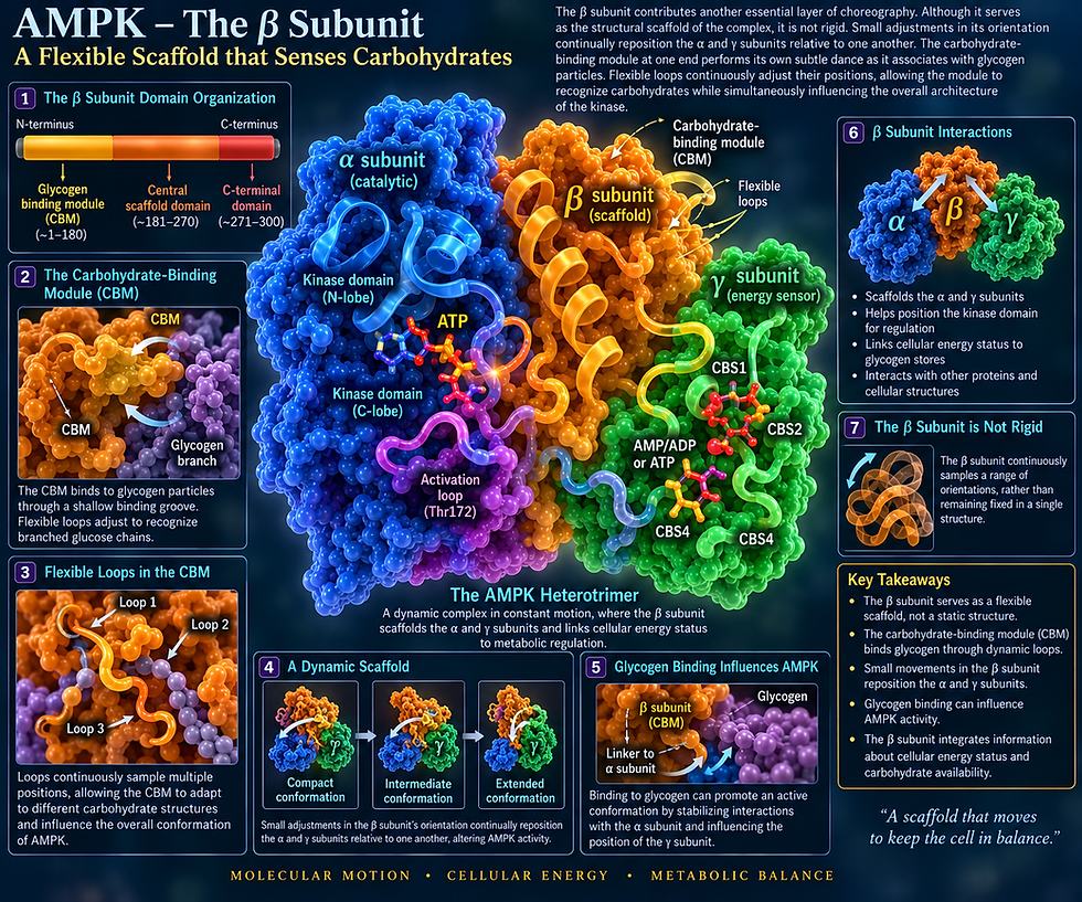
The β subunit contributes another essential layer of choreography. Although it serves as the structural scaffold of the complex, it is not rigid. Small adjustments in its orientation continually reposition the α and γ subunits relative to one another. The carbohydrate-binding module at one end performs its own subtle dance as it associates with glycogen particles. Flexible loops continuously adjust their positions, allowing the module to recognize carbohydrates while simultaneously influencing the overall architecture of the kinase.

Perhaps the most remarkable choreography occurs within the γ subunit, the true energy sensor of the complex. A specialized sequence called CBS domain (Cystathionine-β-synthase) senses cellular energy levels by binding to adenosine nucleotides in AMP, ADP, and ATP. In the γ subunit, four CBS domains assemble into nucleotide-binding pockets that continually monitor the concentrations of AMP, ADP, and ATP.
These domains are not static binding sites. Instead, they repeatedly open, close, twist, and rotate by tiny amounts as nucleotides bind and dissociate. Binding of AMP stabilizes one family of shapes, while ATP favors another. ADP occupies an intermediate position. These seemingly microscopic adjustments propagate through the entire AMPK complex, altering the orientation of the α-linker, repositioning the autoinhibitory domain, and ultimately controlling catalytic activity. The entire kinase responds mechanically to changes in cellular energy through an exquisitely coordinated sequence of molecular movements.

The nucleotide molecules themselves are equally dynamic participants in this dance. ATP entering the catalytic site is constantly changing shape. The adenine ring redistributes its electrons across the aromatic system, subtly altering electrostatic interactions. The ribose sugar rapidly shifts between slightly different puckered conformations. The triphosphate tail bends, twists, and flexes as three negatively charged phosphate groups continually repel one another. Magnesium ions repeatedly bind and release the phosphates, changing ATP's geometry as catalysis approaches. Electron density shifts across the phosphate chain while water molecules reorganize around the active site. ATP is therefore not simply fuel; it is an active participant in a highly dynamic molecular choreography.

AMP, adenosine mono phosphate, the molecule that gives AMPK its name, performs an equally sophisticated dance after binding to the γ subunit. Its phosphate group rotates freely, continuously presenting different oxygen atoms to surrounding amino acids. Its adenine ring samples slightly different orientations while its electron cloud responds instantly to neighboring charges. Binding reorganizes entire networks of hydrogen bonds within the CBS domains, causing small but coordinated structural adjustments throughout the entire complex. A molecule only a few angstroms wide can therefore reshape an enzyme nearly twenty times its size.

Surrounding the protein, water molecules never stop moving. Every exposed surface of AMPK is enveloped by hydration shells that reorganize in trillionths of a second. Hydrogen bonds continually form, break, and reform. Ordered layers of water appear and disappear as domains move relative to one another. These constantly shifting hydration networks help stabilize some shapes while destabilizing others, making water an active participant in AMPK regulation rather than a passive solvent.
Magnesium ions also perform a continual dance. They repeatedly coordinate ATP and ADP, briefly interact with catalytic residues, and exchange water molecules within their hydration shells at astonishing speed. These transient interactions reshape local electrostatic fields and help position phosphate groups for catalysis. Even individual ion contribute to the continuous choreography of energy sensing.
At the fastest timescale, electrons themselves never remain still. Aromatic amino acids redistribute π electrons, charged side chains respond instantly to neighboring ions, hydrogen bonds fluctuate in strength, and electrostatic fields continuously change throughout the protein. These electronic fluctuations occur in femtoseconds, providing the physical foundation upon which every larger conformational movement is built.

The most remarkable feature of AMPK is that all of these motions are coupled together. A single AMP molecule binding to the γ subunit can reorganize hydrogen-bond networks, shift the positions of CBS domains, alter the α-linker, move the autoinhibitory domain, stabilize the activation loop, reposition catalytic residues, reshape ATP within the active site, reorganize surrounding water molecules, and ultimately determine the activity of hundreds of downstream metabolic enzymes. Information flows through the complex as a cascade of coordinated structural movements rather than as a series of isolated chemical events.

This dynamic choreography allows AMPK to integrate an extraordinary variety of signals and interact and communicate with a wide variety of molecules. Changes in ATP production, AMP accumulation, ADP concentration, calcium levels, oxidative stress, nutrient availability, hypoxia, exercise, inflammation, hormones, circadian rhythms, and mitochondrial function all influence different aspects of its structural ensemble. Rather than responding to a single input, AMPK continuously samples an enormous number of shapes, allowing it to calculate the energetic condition of the cell with remarkable sensitivity.
Modern structural biology shows that AMPK is not a static energy sensor but a living dancing, interacting molecule. Every bend of a linker, every rotation of a nucleotide, every rocking domain, every fluctuating hydration shell, every shifting magnesium ion, and every redistribution of electrons contributes to its remarkable ability to interact with a vast number of different molecules in the cell and regulate cellular metabolism.
Like mTOR, its wide-anging functions emerge not from rigid architecture but from dynamic motion and constantly changing interactions and communication among its subunits and with many other molecules. The language through which AMPK senses energy is written in an ongoing choreography of molecular movements that never cease, allowing every cell to balance growth with conservation, abundance with scarcity, and survival with renewal.


