
Smallest Human Virus – Very Dangerous, Very Intelligent
Updated: Aug 25

Hepatitis D virus, also called hepatitis delta virus, is one of the strangest human viruses. Its genome is a tiny circular single-stranded RNA of only about 1,700 nucleotides, and it produces essentially one protein in two forms—the small and large hepatitis delta antigens. It does not encode its own enzyme to copy its RNA as most RNA viruses do. It doesn’t produce its own envelope proteins or most of the machinery normally associated with a virus. Instead, it has an extraordinary ability to use structures and enzymes that already exist in the human liver cell. For production of its new virus particles, it uses envelope proteins supplied by hepatitis B virus.
For size comparisons of dangerous RNA viruses, HIV-1 virus has approximately 9,500 nucleotides with 9 genes in a single strand of RNA that produces 15 proteins. Ebola virus has approximately 18,900 nucleotides with 7 genes in a single strand of RNA that produces 8 proteins. SARS-CoV-2 has 30,000 nucleotides and 11 genes in a single strand of RNA, producing 29 proteins. Influenza has an average of 7,000 nucleotides and 11 genes in 7 or 8 RNA strands, producing12 proteins. Hep D virus accomplishes its entire infectious cycle with one relatively small protein working together with an extraordinarily active RNA molecule.

The journey begins at the surface of a liver cell. The hepatitis D virus particle is surrounded by an envelope containing the three different hepatitis B surface proteins, small, middle, and large. The large surface protein contains a region that bonds to the liver cell receptor. This interaction means that both hepatitis B and D both go right to liver cells.
The hepatitis delta virus attaches to a specific transporter protein on the outer cell membrane of a liver cell. The liver cell's outer membrane pinches inward around the virus, swallowing it whole into an internal vesicle. The vesicle carrying the virus travels deeper into the cell along a pathway toward a vesicle sorting station. Once safely inside this transport station, the viral envelope merges with the vesicle's membrane releasing the ribonuclear protein core of the hepatitis virus into the cytoplasm.
A Viral Genome Arrives in the Cytoplasm
What enters the cytoplasm is not naked RNA. The approximately circular hepatitis D virus RNA with approximately 1700 nucleotides, is extensively covered with a large number of hepatitis delta antigen proteins, forming the ribonuclear protein complex. The antigen protein binds to itself forming structures of two, four, eight, and other amounts of antigen molecules that then cover all parts of the circular RNA genome strand. The virus stays covered while in the cytoplasm. The interactions of the antigen protein with a wide range of cellular molecules is what gives the complex the capacity to move, to be recognized by the cell's transport machinery, and eventually to interact with cellular enzymes. It also protects the RNA genome beneath.
The incoming complex of RNA surrounded by the many antigen proteins, does not remain in the cytoplasm for long. Making copies of hepatitis D virus occurs in the nucleus, yet the virus has no motor of its own for traveling there. Instead, it exploits the cell's nuclear transport system. Hepatitis delta antigen molecules have a particular sequence of amino acids, called nuclear localization signals (NLS), that allow the cell’s nuclear-import machinery to attach to this molecular complex and transport it into the nucleus.
This signal sequence mimics what the cell's own nuclear proteins use to enter. By recognizing this signal sequence, the cell's transport apparatus does the rest, actively carrying the virus genome into the nucleus without hepatitis delta virus needing to expend any energy or build any specialized machinery of its own.

The virus is transported through the large, but well protected, pores in the nuclear membrane by special transporters, called importins, which work along with molecules from the nuclear pore complex for the transport. The antigen molecules surrounding the ribonuclear protein complex produce structures where eight subunits of antigen surround the huge structure. This means that there are eight NLS signals that attract eight importins for the transport. The complex is so large that if there were only one or a few transport molecules, they would not be able to manage pulling it into the pore. By having eight NSL signals attracting eight transport molecules working together, the huge complex is easily moved into the nucleus.
Within a relatively short period after entering the cell, an RNA virus that entered through the cell’s outer membrane has delivered its genome into the cell's nuclear compartment.
Entering the Nucleus and Copying the Virus RNA Genome
Once reaching the nucleus, hepatitis D virus meets its greatest problem and solves it with ingenious wide-ranging manimpulations of the cell's molecules. The virus has an RNA genome but possesses no RNA-dependent polymerase, that is, no RNA copying enzyme, to make the new copies of its genome strand. Almost all other RNA viruses produce their own RNA polymerase, except for hepatitis D virus. The existing polymerase in human cells is used only for copying DNA, not to copy free RNA genomes and the way DNA is copied in the human liver cell involves vastly complex cellular machinery. Hepatitis D virus, nevertheless, convinces this DNA cellular machinery to copy its RNA genome.
The genomic RNA is first copied into a complementary full-length RNA, and this “antigenomic” strand is then used as the template to produce copies of the original genomic strand that will produce the new viruses. The antigenomic RNA is not used except to be the template to make the complementary genome copy, which is used for all virus actions and to produce the messenger RNA for its one protein. Because the virus genome is circular, the polymerase doesn't stop at a defined end but continues around the loop repeatedly, generating a very long RNA strand containing many genome-length repeats linked end to end.
In order to produce a single genome from this long RNA strand with many genomes on it, the RNA folds into a ribozyme that cuts precisely one genome. Then the cell's enzyme attaches the two ends of this cut strand producing the circular hepatitis D virus genome. Copying the antigenic strand to produce the genome strand also produces multiple rolling copies of the genome and it is, also, cut by its ribozyme and the ends are attached making the new circular genome.

During the copying process, the RNA genomic strand is always wrapped by the virus' small antigen proteins and these antigen molecules do not block the cellular copying machinery. In fact, the virus antigen protein is actively involved in recruiting and guiding the cell 's RNA copying polymerase to use the viral RNA as its template. Newly produced genomic RNAs attract newly translated delta antigens from the cytoplasm into the nucleus to form fresh ribonuclear complexes for export. Newly synthesized small hepatitis delta antigen molecules move from the cytoplasm into the nucleus immediately after they are produced by ribosomes using their transport signal (NLS) to stimulate entry.
When the new ribonuclear protein complex returns to the cytoplasm from the nucleus via cell transport systems, it searches for hepatitis B surface antigens so it can envelope itself and escape the cell as a completed virus. If a complex fails to find a hepatitis beta envelope protein in the cytoplasm, the NLS signal for transport to the nucleus on antigen molecules triggers immediately and the complex returns to the nucleus. This continuous shuttling of the ribonuclear protein complex between the cytoplasm and nucleus ensures that the virus maintains an active pool of genomes being copied inside the nucleus while constantly feeding new material to the cytoplasm for full assembly of the virus and release from the cell when possible.
Tricking the DNA Copying Mechanism to Copy RNA
It is the changing shapes of the hepatitis D virus RNA and the protein antigen that fool the cell’s DNA copying enzyme to use this virus RNA that looks like it is a double stranded human DNA. Because the ordinary triggering of DNA copying in the human liver cell is very complex, it takes multiple tricks from hepatitis delta virus to accomplish this.

During the process of copying the RNA genome inside the covered ribonuclear protein complex, the cell’s enzymes force the RNA to unwind so they can read the genetic sequence. Once a new strand of RNA genome has been copied, for a brief moment, before the newly minted circular RNA is bound by fresh delta antigen proteins, the bare RNA instantly snaps into many shapes with multiple levels of folding. Almost immediately, the RNA collapses into a highly stable, double-stranded rod shape. Whenever covered by antigen the RNA shape is circular. Whenever very briefly naked it snaps into a folded rod shape.
The rod shape serves two critical biological functions while inside the cell, even if this folded shape happens only very briefly and transiently. The first function of the rod shape is to trick the cell’s double stranded DNA copying enzyme into using a single strand of RNA that looks like it is double stranded DNA. Because 74% of the rod is base-paired, it appears to be double stranded. The cell enzyme thinks that it is looking at a piece of double-stranded DNA. This allows the virus to hijack a host enzyme that normally only works with double stranded DNA.
The second function of the rigid rod shape is that it distorts the active site of the ribozyme after it has made its cut of the the virus' one genome and this stops the ribozyme from further cutting up the newly closed, functional circular genomes.
In order to proceed making the copies, the cell's DNA copying enzyme must physically attach to hepatitis delta RNA on the loop situated at the very end of the rod formation. It requires specific adaptations to attach to a viral RNA molecule. This is accomplished through a combination of structural mimicry and viral assistance. Specialized regions at the curved "tips" of the RNA rod act as if it is a cellular promotor for copying DNA. Cellular factors bind to this pseudo promotor anchoring the cell polymerase in place.

Still, the cell polymerase cannot load onto the template needed for copying without more help. The polymerase relies heavily on the small viral antigen protein to act as a molecular adapter in 9 of the 12 sub-units of the cell’s polymerase complex. It specifically attaches to the molecular jaw that normally clamps down onto a DNA strand. By loosening this clamp, the small viral antigen protein changes its shape, forcing it to latch tightly onto the viral RNA rod instead of human DNA. Once attached to the rod's tip, the polymerase is able to uncoil the circular RNA surrounded by virus antigen proteins for copying.
The Virus, Actually, Uses Many More Tricks to Fool the DNA Machinery of the Human Liver Cell
The virus, in fact, tricks multiple cellular enzymes for this very complex manipulation of the human liver cell’s vast DNA machinery. It tricks two distinct copying enzymes, polymerase I and polymerase II; it tricks several enzymes related to producing a messenger RNA instead of a full genome; and it tricks the enzyme that changes the nucleotide and amino acid codes from producing small antigen to producing large antigen.
First it tricks the polymerase I copying enzyme. This cell polymerase only copies the incoming virus RNA genome into a complementary antigenic strand of RNA. The antigenic strands is only used as the template to produce copies of the original genome RNA.
Then, it tricks the main polymerase II that copies the complementary antigenic RNA to make the many copies of the original genome to be used for new viruses.
Meanwhile, it also tricks the enzyme that stops the copying after exactly 585 nucleotides to produce the messenger RNA that is used to produce the small protein antigen. These enzymes also process the messenger RNA with a cap on one end and a tail signal molecule on the other that allows the messenger RNA to leave the nucleus and go to a ribosome in the cytoplasm to make small antigen protein.
Later, it tricks the cell enzyme that changes one nucleotide that allows for the production of a long messenger RNA that is used to produce the long antigen, rather than the short antigen. Another cell enzyme prepares this longer messenger RNA for travel to the ribosome. Both of the newly produced antigens, small and large, already have the NLS signal on them that gets them rapidly into the nucleus once they are produced the cytoplasm by ribosomes. Back in the nucleus, the antigens surround a ribonuclear protein complex.
It is quite remarkable that this tiny virus has so many tricks to play on the huge, vastly complex human liver cell.
The Flexible RNA Is Able to Form a Shape Where it Cuts Itself
Another hepatitis D virus trick is being able to do its own cutting, rather than depending on producing an enzyme, as most viruses do.

The virus' cutting ribozyme is based on its own embedded catalytic RNA structure. As the newly synthesized naked genome RNA folds in the instant after it is produced, the ribozyme assumes a precise three-dimensional shape for cutting. It then cuts the long RNA copy at exactly defined positions, generating just one genome on a strand of RNA. The cell’s enzyme then helps by joining the ends of this singular genome together to generate the viruscircular RNA.
To make this cut, the RNA strand automatically folds itself into a tight, rigid pocket. Because of this shape, only one specific chemical bond, which is the target cutting site, gets pushed deep inside the very bottom of the pocket. Right at that exact spot, the RNA chain takes a sharp, awkward twist. This twist pulls the target bond tight and leaves it completely exposed. Waiting at the very bottom of that pocket are specific nucleotides and a magnesium ion. They act like a fixed pair of scissors. It is the unusual ability of the RNA to form many transient shapes that produces the ribozyme.
The hepatitis delta virus ribozyme does not "scout" the whole strand to find where to cut. It is the folding of the RNA sequence itself that forces the cutting mechanism to happen exactly at the junction of each genome unit. The complex folding involves five helical segments that form a rigid, pocket-like active site that brings distant nucleotides into close proximity. The interlocking folds squeeze the helices so closely together that they create a highly shielded deep internal pocket (an active-site cleft) for lightning-fast self-cutting.
In order to free a complete genome from the long strand with many genomes end to end, it must have both ends of the strand cut. Because a ribozyme sequence is part of each genome in the long strand, each will cut one end of its genome with its ribozyme and the other end will be a cut by the next genome's ribozyme. Either simultaneously or sequentially, both ribozyme sequences fold into their active structures and perform a self-cutting reaction that severs the bonds, establishing the exact, clean start and end sites of the individual genome. This linear unit is then connected into a circle by cell enzymes to form the final, infectious circular virus genome.
Hepatitis delta virus therefore carries enzymatic information directly in the changing shapes of its RNA. The genome is simultaneously genetic information, structural scaffold, polymerase template, and a catalytic molecule.
Making the Small Hepatitis Delta Antigen

Hepatitis delta virus produces its antigen protein from its polymerase copying cycle. A messenger RNA is generated in the nucleus that is processed by the liver cell's normal messenger RNA machinery and then exported to the cytoplasm. Cellular ribosomes in the cytoplasm produce the small hepatitis delta antigen protein, which is 195 amino acids long.
The small hepatitis delta antigen is one of the most striking examples of an intrinsically disordered viral protein, that is, a protein strand that constantly changes shapes, appearing to be dancing. Although the antigen is small, it performs dozens of functions by constantly changing its shape as it interacts with viral RNA and host proteins. In fact, 70% of the antigen is intrinsically disordered, with no more than about 30% adopting stable α-helical structures under normal conditions. This percentage changes depending on what molecule it is interacting with.
The changing structures are not random but continually shift among thousands of shapes, each exposing or hiding different binding surfaces, allowing the same protein to interact with viral RNA, cell RNA polymerase, nuclear chromatin proteins, protein transcription factors, nuclear transport machinery, RNA-processing proteins, and many other cellular partners.
More than one hundred cell proteins have been identified as interacting partners of the small antigen, an extraordinary number for such a small viral protein. A small list of its functions include forming the viral ribonuclear protein complex; multiple ways of promoting the rolling circular copying of the genome; helping to recruit and regulate host RNA polymerase to copy the viral RNA; establishing the complex that generates the cap and tail of the messenger RNA to produce the antigen; serving as scaffold for cell complexes; and many others.
The antigen’s many shapes and therefore its many functions are increased even further because it is tagged with added molecules by the cell in multiple ways. These chemical tags include adding a phosphate, a methyl group, an acetyl group, and others. Depending on its binding partner and its pattern of chemical modifications, the antigen can act as an RNA-binding protein, a scaffold, a regulator of copying, and a chromatin-associated factor. These many different structural forms allow an exceptionally small viral genome to orchestrate a remarkably complex infection cycle
The hepatitis delta virus antigen is essential for efficient RNA copying. It participates in the organization of viral ribonuclear protein, interacts with cell proteins, interacts with the polymerase enzyme while making copies, and helps establish the molecular environment required for continuing RNA synthesis.
The Decision of Producing A Messenger RNA Versus a Whole Circular RNA Genome
When the polymerase latches onto the circular virus genome RNA, it makes a choice to either make a messenger RNA to produce antigen or to make the full circular genome, based on the state of the virus lifecycle. The primary "switch" that decides whether to make messenger RNA or a circular genome is the effect of the accumulation of the antigen proteins and modifications in function produced by the antigen proteins.
To produce messenger RNA, the polymerase starts at a specific promoter site on the RNA template and stops at a dedicated signal site, instead of going around the entire circular RNA. This produces a short, linear, partial copy, approximately 800 nucleotides long. Instead of using the viral ribozyme to cut out the messenger RNA, the virus hijacks the cell's own protein machinery to cut the strand. Cell enzymes recognize the signal for messenger RNA, slice the two ends of the linear RNA strand.
Cell enzymes, also, immediately paste a cap on one end of the messenger RNA strand and a special tail onto its other end, which is a signal that allows it to travel to the cytoplasm to find a ribosome where the antigen protein is produced. This tail is approximately 200 adenosine nucleotides and protects the mRNA from attacks, helps export it from the nucleus, helps with producing proteins, and determines how long it will live.

To make full circular genome copies, the virus must suppress the messenger RNA pathway. Whenever there are free small antigens nearby that are not bound to a ribonuclear protein complex, they bind directly to the polymerase enzyme. When small antigen binds to the polymerase, it acts as an elongation factor, that is, a message to ignore the stop signal that is needed to make a messenger RNA, and to keep going. The result is that when a lot of antigen is present in the nucleus and attached to the polymerase, it drives the copying continuously around the circular template over and over, generating the massive, multi-genome linear strands. The built-in self-cutting RNA ribozyme slices this massive strand into precise, full-length genome pieces, which are then glued together into complete, circular genome copies.
Producing a messenger RNA versus a total circular genome is part of a hepatitis virus lifecycle in three phases—making small antigens, focusing on full circular genomes, and making large antigens. When the virus ribonuclear protein complex first enters the cell nucleus, there are few free floating antigen molecules, with most of the viral antigen proteins surrounding the ribonuclear protein complex that just entered the nucleus. There are a few free floating antigen molecules available to modify and work with the cell polymerase machinery. With only a few antigens present, the polymerase obeys the stop signal to produce messenger RNAs.
The messenger RNA used to make antigen protein travels to the cytoplasm where it produces many antigen copies, each with the NLS nuclear import signal so it can go very easily to to the nucleus. The antigen proteins accumulate in the nucleus where they physically bind to the cell’s RNA polymerase. As the pool of small antigen proteins grows, which are bound to the polymerase, the polymerase switches entirely from making messenger RNA for proteins to copying the full circular genetic material. With the attached small antigen, the polymerase ignores the stop signals to make messenger RNA and keeps going around the circle copying the genome many times. Then, this long strand is cut by the ribozymes into individual genomes, which are bound at both ends to make circular genomes.
After producing many of these circular viral genomes, there is an accumulation of antigenic genomes, and this produces a switch triggering the third phase in the life cycle. A particular cell enzyme modifies the viral RNA, causing the polymerase to switch from making large circular genomes, and to start producing messenger RNA again. But, this time because of the modification that was made in the RNA, it produces messenger RNA for the larger version of its antigen protein. This switch to production of the large antigen, the third phase of the virus life cycle, is accomplished by a special cell enzyme which edits a nucleotide on the hepatitis delta virus RNA.
This altered nucleotide occurs on the antigenic comlementary strand, so that when the next genome strand is produced from this altered (mutated) antigenic strand, the genomic strand starts producing messenger RNA for the large antigen. The effect of more large antigens is to inhibit further RNA copying altogether. This signal directs the newly formed circular genomes to leave the nucleus to be packaged into viral envelopes so they leave the cell and infect new targets.

One RNA Letter Changes the Strategy and the Phase of the Life Cycle
As virus infection of the cell progresses, hepatitis delta virus performs one of its most ingenious regulatory switches. It manipulates one of the cell's enzymes to trigger the change from producing full circular hepatitis D genomes to producing the large antigen protein and changing its focus to start assembling the full virus and leave the cell.
The critical enzyme is called ADAR1 (stands for Adenosine Deaminase Acting on RNA 1) and this attacks a particular adenosine nucleotide on the hepatitis D virus RNA, when it is able to reach it. In fact, this enzyme is always attempting to attack the adenosine, but can only reach it when hepatitis D ribonuclear protein is in its naked rod like shape and exposed.
Most of the time, the genomic strand of RNA is completely covered by small antigen molecules and therefore ADAR1 can't reach it. This covering protects the genome from cell enzymes that would destroy it. The covering of small antigen molecules also help with the process of partial uncoating of the naked RNA to proceed with copying. When the small antigen binds tightly to the RNA, it stretches, stabilizes, and partially opens up the RNA, preventing it from folding into its tight, double-stranded rod shape. When small antigen covers the ribonuclear protein complex, the site for editing is hidden.
The antigenic strand is not as covered and protected as well with antigen protein as the genomic strand. This leaves some regions exposed when it forms the rod shape. In fact, at the moment right after copying, the RNA immediately flips into the rod like shape, before small antigen starts to cover it. It is at this moment that the adenosine is most exposed to ADAR1. After the entire copying process has proceeded for a time, there are more and more antigenic genomes accumulating and this allows the editing site to be exposed more and more often.

When these sites on the rod-like form of the antigenic genome are exposed, ADAR1 attacks the particular adenosine at the exposed site and converts an adenosine nucleotide of the hepatitis delta virus to an inosine nucleotide. Inosine is not a usual nucleotide used in RNA or DNA. At the ribosome, the inosine is interpreted as if it is a guanosine nucleotide, rather than the original adenosine.
When the RNA strands flips transiently into its rigid rod shape it exposes the vital loop that is attacked by ADAR1 and in this loop the nucleotides are UAG, that is uracil, adenosine, and guanosine. When ADAR1 is able to attack at that instant ADAR1 latches onto the RNA loop at the end of the rod and changes the loop's shape so that the reaction can occur. This reaction takes off the NH2 group attached to the adenosine ring (hence "de-aminate" in its name) and replaces it with an oxygen. This is what changes the adenosine to inosine.
So, now the nucleotides read UIG (uracil, inosine, guanosine) instead of UAG. When this antigenic strand becomes a template for polymerase to make a genomic strand, the inosine is still there. But, since it is not a regular letter, it is read by the polymerase as a G, for guanine (one of the normal letters). So, actually the new edit changes UAG to UGG. That is, it changes the three nucleotides uracil, adenosine, guanine to uracil, guanine, guanine.
UAG stands for a stop signal and UGG stands for the amino acid tryptophan when making the antigen protein.
When this strand is converted back into a genomic template, the sequence becomes CCA (the exact complement of UGG). This means the virus now possesses a mutated genomic template that permanently encodes a tryptophan amino acid instead of a stop signal. When cell RNA polymerase II binds to this new, edited genomic template to produce a messenger RNA of the antigen, it passes right through the newly created tryptophan (UGG) without stopping.
The consequence of this one nucleotide change is dramatic. The change in nucleotides affects the stop signal that normally terminates synthesis of the small antigen. The signal to stop is changed from “stop” to the amino acid tryptophan and therefore a new larger antigen protein is produced with an additional 19 amino acids, until it reaches the next stop signal. This remarkable event produces the large antigen protein which is 214 amino acids long. The messenger RNA that is produced for the large antigen also is processed by the cell enzymes adding a cap on one side and a tail of many adenosines to aid its trip to the cytoplasm and the ribosome to produce large antigen proteins.
This single nucleotide RNA-editing event changes the entire viral program. Early in infection, the virus cycle favored messenger RNA for the small antigen. Then, with plenty of small antigen available to cover the ribonuclear protein complex, the cycle favored production of the full circular genome. Now, this change triggers the third phase of the life cycle, which is production of the large antigen. The effect of more large antigen is to suppress copying of the genome and instead to promote assembly of new viral particles to leave the cell. Hepatitis delta virus has effectively converted a cell RNA-editing enzyme into a developmental switch.
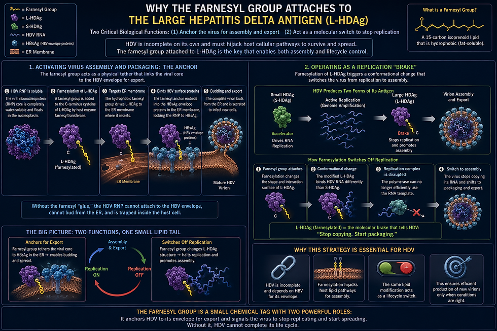
The Large Antigen, Adding a Lipid Group, and More Tricks
The large antigen protein is also mostly intrinsically disordered, retaining the flexible, constantly moving, interaction surfaces that characterize the delta small antigens. This flexibility enables rapid transitions between interactions with viral RNA, transport proteins, membranes, and hepatitis beta virus envelope proteins.
The extra 19 amino acids at the end of the large antigen provides several more tricks, with multiple vital new sequences of amino acids, one that is modified by yet another cell enzyme. This cell enzyme covalently binds a 15-carbon lipid molecule to the cysteine amino acid at the end of the large antigen. This farnesyl lipid group is highly greasy and hydrophobic. It acts like a physical anchor into fatty membranes that allows the viral ribonuclear protein core to insert into cell membranes where it meets hepatitis beta surface antigens embedded in the membranes at the endoplasmic reticulum.
The farnesylation changes the large antigen behavior in muliptle important ways. One is that it forces the antigen complexes out of the nuclear machinery where replication happens, moving them toward areas where virus assembly takes place. Secondly, once farnesylated, the large antigen protein binds to host cell RNA polymerase II and blocks it from elongating the viral RNA strand.
This chemical modification has another enormous consequence. By attaching this lipid group to the large antigen, the virus changes the interaction properties of its entire ribonuclear protein complex and gives it the ability to associate effectively with the hepatitis B virus surface proteins, which becomes the envelope for the new hepatitis delta viruses. We already noted at the beginning of this journey that it is these hepatitis B molecules that are so vital in allowing the virus to first enter the cell. Placing them on the new delta viruses allows them to enter new cells once they leave the cell.
The two delta antigens therefore represent different phases of the same viral strategy. The small antigen strongly supports copying. The large antigen shifts the system toward assembly of the new virus and leaving the cell.

The Same Cellular Polymerase Does Three Unique, Different, Unusual Processes
For clarity, this is a summary of material already presented about possibly hepatitis D virus' most remarkable behavior. In the cell, usually three different polymerases produce antigenome, genome, and messenger RNA.
For hepatitis D virus, the cell’s RNA polymerase II (Pol II) is manipulated into producing all three major RNA products in the virus life cycle. It does this even though this polymerase does not normally work with RNA, but only DNA. This particular cellular polymerase normally produces messenger RNA from DNA, not genomes and antigenomes.
This polymerase produces these three results in different contexts, three different manipulations by the virus, with different initiation mechanisms and different termination mechanisms. The remarkable aspect is that the same cellular enzyme is effectively repurposed to perform three quite different jobs.
RNA polymerase II produces the multimeric antigenome of the virus. Instead of making one copy and stopping, Pol II continues traveling around the circular template repeatedly, producing a long linear multimeric antigenome containing many genome-length copies joined end to end. This genome has no cap, has no poly(Adenosine) tail, is not mRNA, is later cut by the hepatitis D virus ribozyme, is joined into circular antigenomes.
The same polymerase produces the multimeric genomic RNA of the virus. The newly circularized antigenome becomes the template for the genomic RNA. Again, RNA polymerase II repeatedly circles this template, producing long multimeric genomic RNA. Again: no cap, no poly(Adenosine) tail, ribozyme cleavage, joined into new circular genomes.
The same polymerase also produces the antigen messenger RNA. Instead of continuous rolling-circle transcription, it initiates at an mRNA-producing region on the genomic RNA and synthesizes only the antigen messenger RNA.This RNA is processed by the cell exactly like a normal cellular messenger RNA. It receives a cap on one end, receives a poly(Adenosine) tail on the other, is exported from the nucleus, and used to produce proteins by cell ribosomes.
Initially this messenger RNA producers small antigen. Later, after ADAR1 edits the antigenome and changes a stop to a tryptophan amino acid, the same messenger RNA region ultimately produces large antigen with its additional 19 amino acids.
The polymerase itself is essentially the same enzyme in all three processes, but its associated factors and RNA template determine what kind of RNA is produced. For rolling-circle replication Pol II ignores normal gene boundaries. It circles the RNA template repeatedly. No capping or polyadenylation occurs.
For messenger RNA synthesis Pol II recruits, the normal host messenger RNA-processing machinery. The RNA produced is capped almost immediately. Cutting and tail factors recognize the appropriate termination signals. The process stops after a single messenger RNA rather than continuing around the circle.
The difference lies in the molecular complex assembled on the RNA, including cell genome copying factors, RNA structure, associated antigen proteins, and RNA-processing proteins.
This ability to use a single host polymerase for all three is one of the most extraordinary examples of molecular adaptability in virology. Perhaps the most astonishing aspect of hepatitis D virus is that it persuades the cell's RNA polymerase II—an enzyme evolved exclusively to transcribe DNA into messenger RNA—to perform two entirely foreign tasks, : copying RNA into RNA to generate both antigenomes and genomes, while still carrying out its normal function of producing the viral messenger RNA.
Leaving the Nucleus and Meeting Hepatitis B at the Endoplasmic Reticulum
After sufficient new genomic virus RNA has accumulated and associated with antigen forming new ribonuclear complexes in the nucleus, these must now travel to the cytoplasm where final assembly takes place.
Within the extra 19 amino acids, the large antigens have two other amino acid sequences that are special signals. One is the a amino acid sequence called the Nuclear Export Signal (NES), which recruits cell transport complexes to ferry the entire viral core out of the nucleus and into the cytoplasm.
This transit places the ribonuclear protein complex in the cytoplasm just at the endoplasmic reticulum, where it meets hepatitis beta virus. Another signal sequence of amino acids is called the "clathrin box" that also tricks the cell into wrapping the virus complex into transport vesicles destined eventually for the exit. This sequence allows proteins to bind directly to the large structural proteins (clathrin heavy chains) that build transport vesicles inside the cell to be used by the secretory transport system.

Nuclear export of hepatitis delta virus ribonuclear complexes involves viral RNA, delta antigen, and the cell’s nuclear-export machinery. Newly produced genomes move from the nuclear center of copying into the cytoplasm where they are taken up in the elaborate secretory system of transport around the cell.
The secretory system is a highly organized, interconnected network of organelles responsible for producing, modifying, sorting, and transporting proteins and lipids to specific cellular compartments. It operates like a specialized factory assembly line, moving cargo with transport vesicles through the entire cell including releasing these vesicles out of the cell.
However, at this point, moving into the cytoplasm, the virus faces another major problem. It cannot manufacture its own viral capsid or membrane envelope as most viruses do. This is where hepatitis beta virus becomes essential for the hepatitis delta virus life cycle.
When the cell produces hepatitis beta virus surface antigens, they are placed into membranes of the endoplasmic reticulum and associated secretory transport compartments. It is the farnesyl lipid group attached to the hepatitis delta virus large antigen that acts as a physical hydrophobic anchor that inserts directly into the endoplasmic reticulum membrane. This positions the hepatitis delta viral core exactly where it can bind to hepatitis B surface antigens, which become the delta virus envelope protein. The hepatitis delta virus genome and delta antigens thus become bound to a completely different virus's membrane proteins.
The relationship is highly specific. Hepatitis delta virus does not need hepatitis beta virus to copy its RNA or manufacture antigens. It needs hepatitis beta virus to provide the envelope machinery required to assemble and release the virus infectious particles out of the cell with the ability of efficiently entering another liver cell.

Building a New Virus and Leaving the Cell
At endoplasmic reticulum membranes, the hepatitis delta virus ribonuclear protein becomes enclosed by an envelope containing hepatitis beta virus surface proteins in a membrane cover. The mature hepatitis delta virus particle is therefore a biological composite. Inside is the delta circular RNA associated with its own delta antigens, both small and large. Outside is a lipid membrane decorated largely with proteins encoded by beta.
This arrangement solves several problems simultaneously. The beta-derived envelope protects the ribonuclear protein, provides a mechanism for budding from the endoplasmic reticulum into the cellular secretory transport pathway, and supplies the receptor-binding machinery necessary to infect the next liver cell.
With the beta antigens enmeshed with membrane covering the hepatitis D ribonuclear protein complex, it becomes an enveloped virus particle sent into the secretory transit system. It is the clathrin box signal that helps package the virus into a particle that can be transported in the secretory pathway. The secretory pathway takes it from the endoplasmic reticulum through the Golgi apparatus, following the same route ordinary secreted membranes and membrane proteins take, and is ultimately released from the liver cell. The release mechanism, called exocytosis, is the same that the cell uses for all its lipid covered vesicles, that are sent into the blood as messages to other cells. The fully developed virus particle with its envelop are then released from the liver cell where they can encounter neighboring or distant liver cells.

The hepatitis beta virus-derived envelope now performs another role. Its large surface protein can recognize the receptor on another liver cell, beginning the entire sequence again: attachment, entry, cytoplasmic release, nuclear import, RNA copying, ribozyme cleavage, antigen production, RNA editing, attachment of lipids, nuclear export, envelope acquisition, assembly, and secretion.
An Extraordinary Density of Information and Intelligence
What makes hepatitis delta virus remarkable is not simply its small size but how much biological organization it obtains from so little genetic material. It hijacks a liver cell’s own machinery, and hepatitis beta virus, so thoroughly that it needs almost nothing of its own beyond that single gene product and a strand of circular RNA. This stripped-down virus compensates for what it lacks by borrowing everything else. It the constantly moving, dancing, disordered behavior of both the antigen protein, and the RNA molecule, that enable the vastly complex transient shapes that produce all of these remarkable intelligent actions.
With a tiny amount of RNA and protein, this virus manipulates the vastly more complex human cell. With all that it accomplishes, how can this virus not be considered intelligent?


